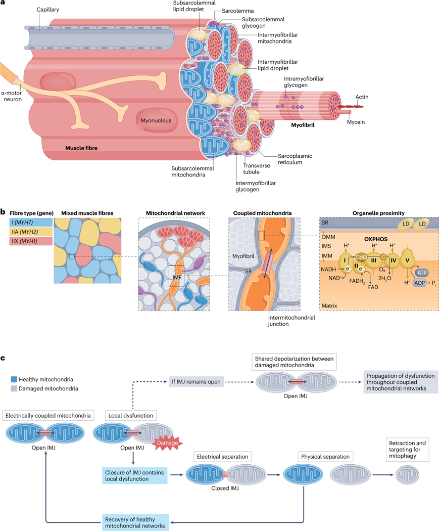
Fig. 1 |. Skeletal muscle fibre ultrastructure.
a, Location of mitochondrial subpopulations and energy stores in muscle fibres. Skeletal muscle is composed of layers of connective tissue and fascicles (also known as muscle bundles). Fascicles contain organized arrangements of individual syncytial muscle fibres, each covered by an endomysium, or basal lamina, which is anchored to the fibre membrane (also known as the sarcolemma). Muscle stem cells, termed ‘satellite cells’, reside within this sarcolemma-basal lamina ‘niche’ (Supplementary Fig. 3). Specialized components, such as sodium/potassium pumps (Na+/K+-ATPase), triads (consisting of transverse tubule and sarcoplasmic reticulum (SR)) and proteins of the myofibrils (long arrangements of connected sarcomeres) enable fibre contraction through the process of excitation–contraction coupling and sliding filament theory (Supplementary Fig. 2). Free ATP in muscle is limited69,70, and fibres possess additional energy depots to maintain contractile activity, including creatine phosphate, glycogen and intramyocellular lipids (Box 3). Glycogen granules are nonuniformly distributed between intramyofibrillar, intermyofibrillar and subsarcolemmal pools35,267,268. Alternatively, intramyocellular lipids are stored in lipid droplets (LDs) found predominantly at central (intermyofibrillar) but also peripheral (subsarcolemmal) regions within healthy muscle fibres76,78. During submaximal54 and longer-duration high-intensity interval55 exercise most ATP in muscle is regenerated by mitochondrial oxidative phosphorylation (OXPHOS) (see the section ‘Acute exercise muscle metabolism’) (Fig. 2). b, Spatial distribution of the mitochondrial reticulum within muscle fibres. Human muscle comprises three main fibre types14,21,22,24,36, type I (marked by MYH7 expression), type IIA (with MYH2 expression) and type IIX (expressing MYH1). Differences in mitochondrial protein content14,21 and mitochondrial network configuration56,60 between fibre types directly impacts muscle metabolism and function. Muscle mitochondria form an interconnected reticulum56–60 that enables swift and efficient distribution of potential energy from subsarcolemmal (also known as peripheral) mitochondria to intermyofibrillar mitochondria (IMF), deep within the fibre56–58. The positioning of mitochondria in the intermyofibrillar space influences the structure of adjacent sarcomeres, resulting in variable cross-sectional areas and myofilament spacing at different regions across the sarcomere length53. The branching morphology of IMF also accommodates functional interactions with nearby cellular components, such as the sarcoplasmic reticulum and intermyofibrillar lipid droplets53,56,58. In oxidative mouse muscle, ~20% of all IMF are connected to lipid droplets, which may facilitate efficient ATP production and distribution56. c, Adjacent mitochondria form networks and share energy potential through the intermitochondrial junction (IMJ). Analogous to circuit breakers, intermitochondrial junctions split the reticulum into smaller subnetworks, permitting swift separation of defective mitochondria before their removal through mitophagy58. In this way, intermitochondrial junctions provide a dynamic layer of quality control, rapidly rewiring the mitochondrial reticulum through healthy network components to sustain muscle function58. C, cytochrome c; CoQ, coenzyme Q; IMM, inner mitochondrial membrane; IMS, intermembrane space; OMM, outer mitochondrial membrane.

