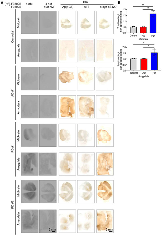
Figure 4. [18F]-F0502B binding affinity and selectivity to different human brain slides.
(A) Autoradiographic labeling of adjacent brain sections from patients with [18F]-F0502B (4 nM). Total binding of [18F]-F0502B was markedly abolished by the addition of non-radioactive F0502B (400 nM), except for the nonspecific (NS) labeling of white matter (left 2 lanes). Scale bars, 5 mm. The serial sections of each group were immunostained with antibodies against Aβ, p-Tau, and pS129 α-Syn (right 3 lanes). PD patient brains displayed specific [18F]-F0502B signals coupled with demonstrable pS129 staining. Scale bars, 5 mm.
(B) Quantification of [18F]-F0502B binding in human brain slices. The total mean signal intensity for specific brain regions was normalized to the nonspecific signal intensity of the same region. Error bars represent the mean ± SEM. Statistical significance was determined using a two-way ANOVA followed by post hoc Bonferroni test for multiple group comparison. Data represent three independent experiments using 3 controls, 3 AD patients, and 3 PD patients in the midbrain and amygdala. *p < 0.05, **p < 0.01.

