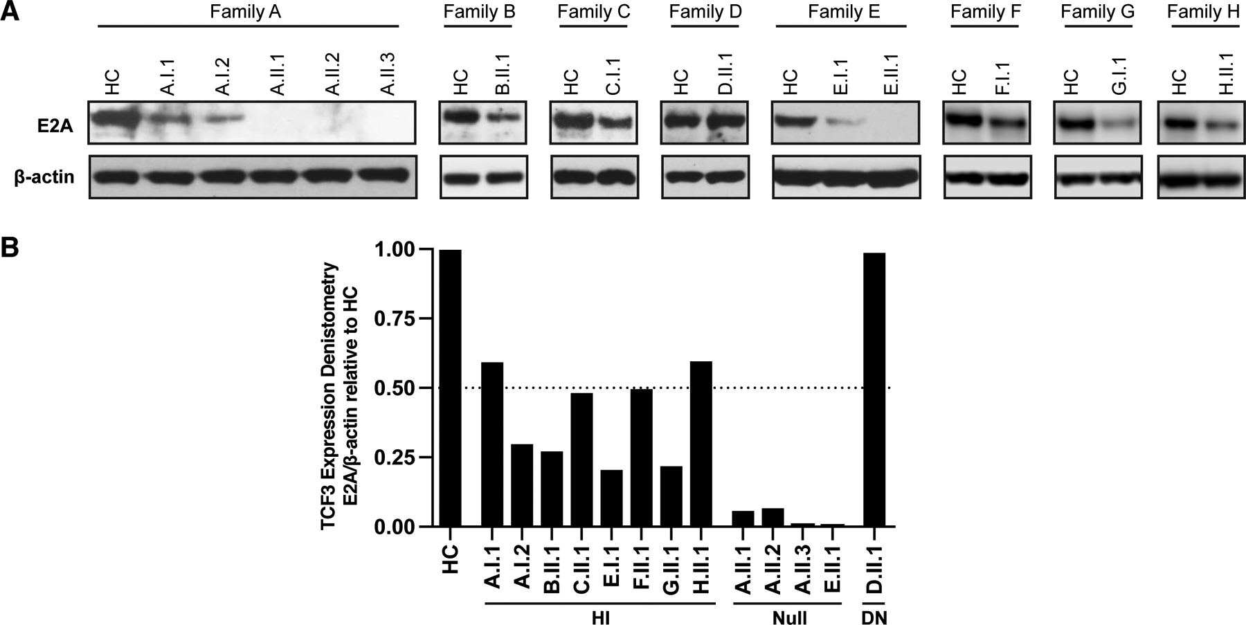
Figure 2:

TCF3/E2A protein expression in T cell blasts. (A) TCF3/E2A protein expression from T cells blasts from healthy controls (HC) and TCF3-mutated individuals. PBMCs were stimulated with anti-CD3/anti-CD28 (1μg/ml each) plus IL-2 (20ng/ml) for 7–12 days followed by protein lysate preparation and immunoblotting with a TCF3/E2A antibody that recognizes both E12 and E47 isoforms. Data shown is representative of two independent experiments. (B) Densitometry of E2A protein expression normalized to β-actin and relative to the HC samples run on the same gel, is shown for TCF3 haploinsufficient (HI), null, and dominant negative (DN) individuals.
