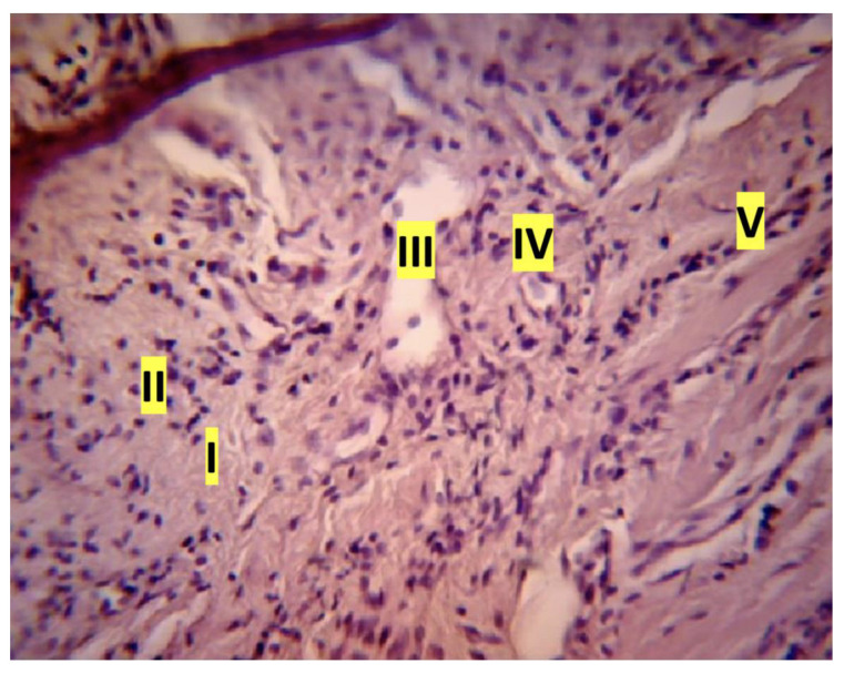
Figure 3.
Exudative inflammation of rat gingival tissue: macrophage reaction in the mucosal lamina, hematoxylin and eosin (magnification ×100): I—edema-swelling, disorientation of collagen fibers; II—the beginning of leukocyte infiltration of the tissue; III—dilatation of the venule with parietal standing of macrophages; IV—macrophage in the cross section of the capillary; V—accumulation of leukocytes in the capillaries (longitudinal section of the vessel)).

