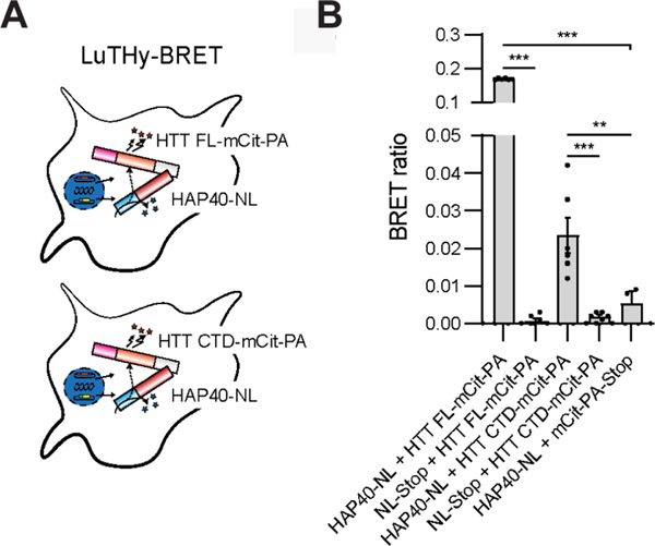
Figure 6: LuTHy assay shows interaction of HTT full-length (FL) and its C-terminal domain (CTD) with HAP40 in live cells.
A, Graphical illustration of the LuTHy-BRET assay performed. HTT FL or HTT CTD were expressed as mCitrine-Protein A (mCit-PA)- and HAP40 as NanoLuc luciferase (NL)-tagged fusion proteins in HEK293 cells. After expression for 48 h and addition of luciferase substrate, BRET was quantified from live cells. B, BRET ratios between HAP40-NL and HTT FL-mCit-PA and HTT CTD-mCit-PA. As a control, NL only was co-transfected with the HTT acceptor constructs, respectively, as well as PA-mCit only with the HAP40-NL donor construct. HAP40-NL co-expressed with HTT FL-mCit-PA and HTT CTD-mCit-PA showed significantly increased BRET ratios compared to controls, respectively. Bars represent means ± SEM from two independent experiments performed in triplicate. One-way ANOVA with Tukey’s multiple comparisons test, **p < 0.002, *** p < 0.001.

