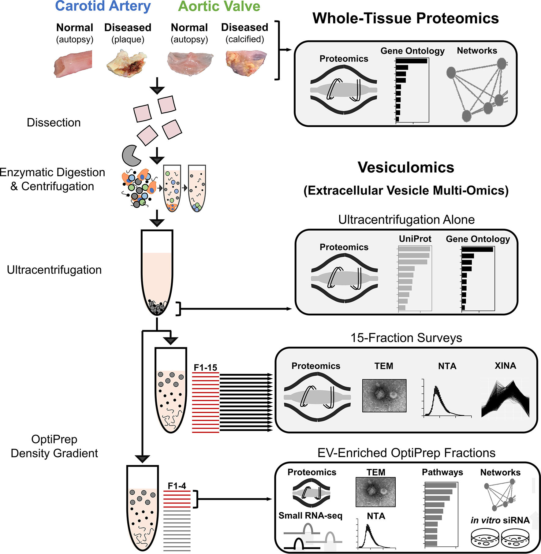
Figure 1: Experimental Overview – Isolation and Analysis of Cardiovascular Tissue-Entrapped Extracellular Vesicles.

Label-free proteomics was conducted on whole-tissue samples of human normal carotid arteries, diseased carotid artery atherosclerotic plaques (from carotid endarterectomies), normal aortic valves, and diseased calcified aortic valves (from valve replacement surgeries for aortic valve stenosis). These sample types also underwent enzymatic digestion and serial low- and high-speed centrifugation. The high-speed supernatant then underwent ultracentrifugation to wash/pellet EVs. 15-fraction survey experiments utilized OptiPrep density gradient separation in concert with mass spectrometry, transmission electron microscopy (TEM), nanoparticle tracking analysis (NTA), and coabundance profiling (XINA) to identify which fractions were enriched in EVs and which contained non-EV contaminants. Enrichment performance was compared to that of ultracentrifugation alone by performing mass spectrometry on the pellet resulting from ultracentrifuged tissue digests. Tissue EV cargoes were then assessed by pooling EV-enriched OptiPrep fractions (F1–4) together and employing mass spectrometry, transcriptomics, TEM, NTA, and bioinformatics approaches to interrogate and classify these vesicles.
