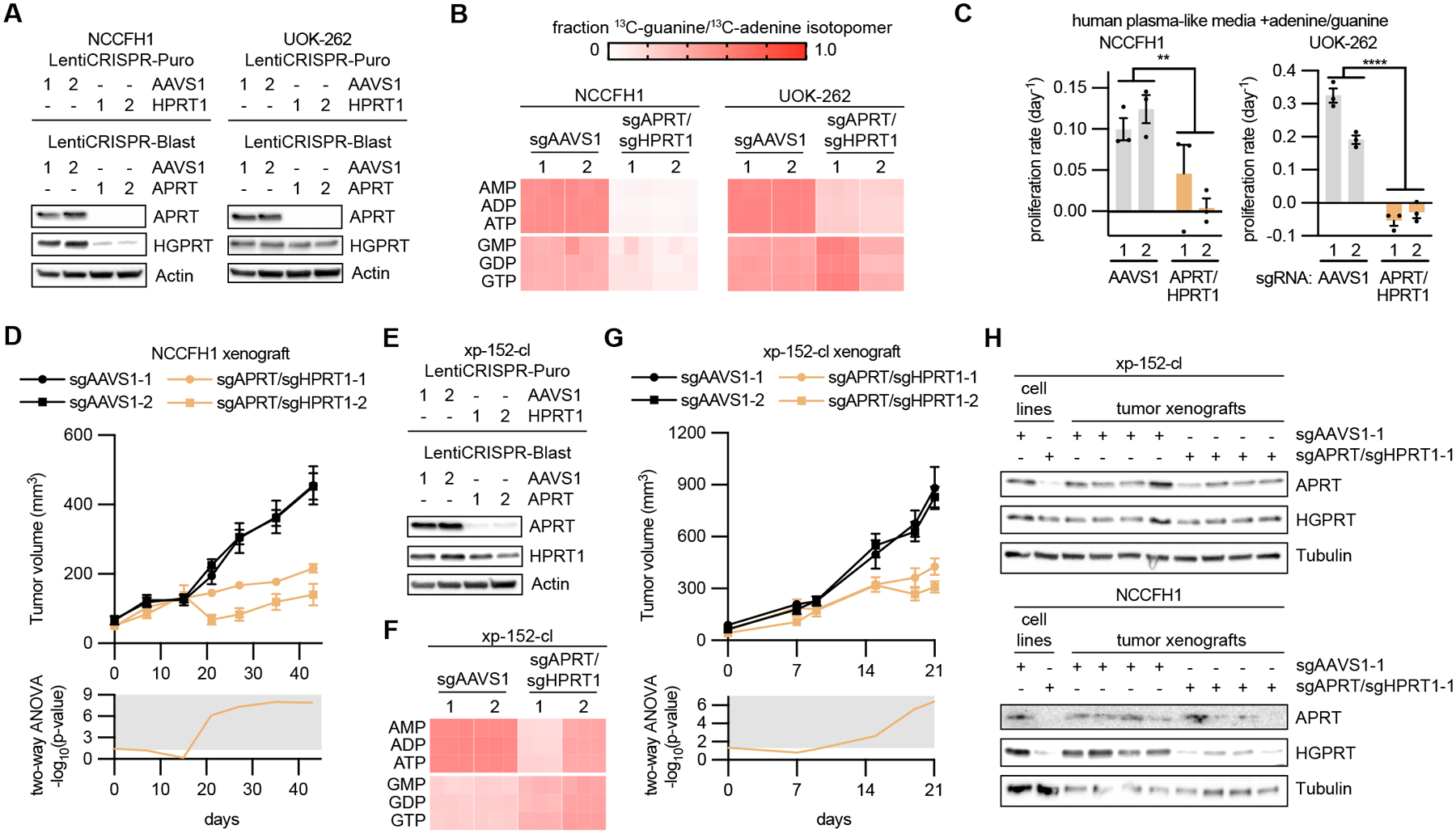
Fig 6: Purine salvage pathway enzymes promote HLRCC tumor growth.

(A) Immunoblots evaluating APRT and HGPRT expression in NCCFH1 and UOK-262 cells infected with LentiCRISPR-Puro and LentiCRISPR-Blast containing sgRNAs targeting AAVS1 (control), APRT, and HPRT1. (B) Fraction of m+1 purine nucleotide isotopologues resulting from 6 hour treatment with 50 μM 8-13C-adenine and 50 μM 8-13C-guanine. (C) Proliferation rates in cells expressing sgAAVS1 or sgAPRT/sgHPRT1 at treated with human plasma-like media supplemented with 50 μM adenine and 50 μM guanine. (D) Volume of NCCFH1 tumor xenografts expressing sgAAVS1 or sgAPRT/sgHPRT1 as determined by caliper measurements (n = 10). Two-way ANOVA was performed for each time point and -log10(p-value) is plotted below, with significant values (p ≤ 0.05) falling into the gray region. (E) Immunoblots evaluating the expression of APRT and HGPRT in xp-152-cl cells infected with LentiCRISPR-Puro and LentiCRISPR-Blast containing sgRNAs targeting AAVS1 (control), APRT, and HPRT1. (F) Fraction of m+1 purine nucleotide isotopologues resulting from 6 hour treatment with 50 μM 8-13C-adenine and 50 μM 8-13C-guanine. (G) Volume of xp-152-cl tumor xenografts expressing sgAAVS1 or sgAPRT/sgHPRT1 as determined by caliper measurements (n = 8). Two-way ANOVA was performed for each time point and -log10(p-value) is plotted below, with significant values (p ≤ 0.05) falling into the gray region. (H) Immunoblots evaluating the expression of APRT and HGPRT in lysates from NCCFH1 and xp-152-cl cell lines and corresponding tumor xenograft lysates.
