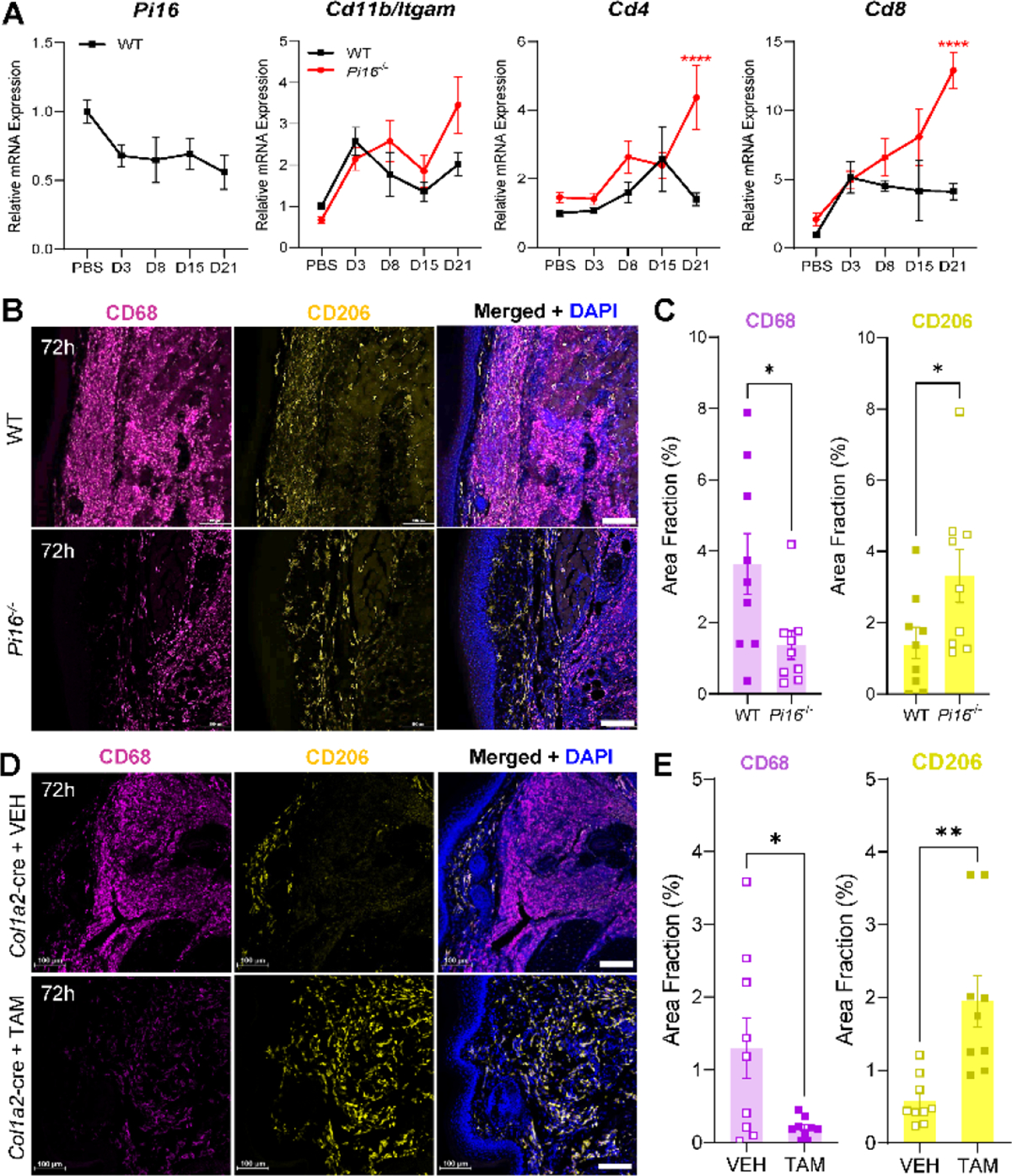
Fig. 12.

PI16 deletion is associated with reduced macrophage infiltration of hindpaw skin following CFA. (A) qPCR shows no significant expression changes in Pi16 in WT skin, or Itgam/CD11b in female Pi16−/− versus WT hindpaw skin. Significant increases in expression of T-cell markers CD4 and CD8 occur only at late-stage timepoints. (B) Hindpaw skin shows significantly reduced CD68 (magenta) immunoreactivity along with an increase in the area fraction of CD206hi (yellow) cells in female Pi16−/− mice. DAPI: blue. Scale bar: 100 μm. (D) Representative IHC in vehicle (VEH) and tamoxifen-induced fibroblast PI16 knockout (Col1a2-cre) plantar hindpaw skin, 3 days after intraplantar CFA. (E) Significant decrease in CD68+ cell density in skin from mice with fibroblast-specific deletion of PI16. Significant increase in CD206hi cell density in skin from mice with fibroblast-specific deletion of PI16. Student’s T-test: *P<0.05, **P<0.01, Student’s T-test.
