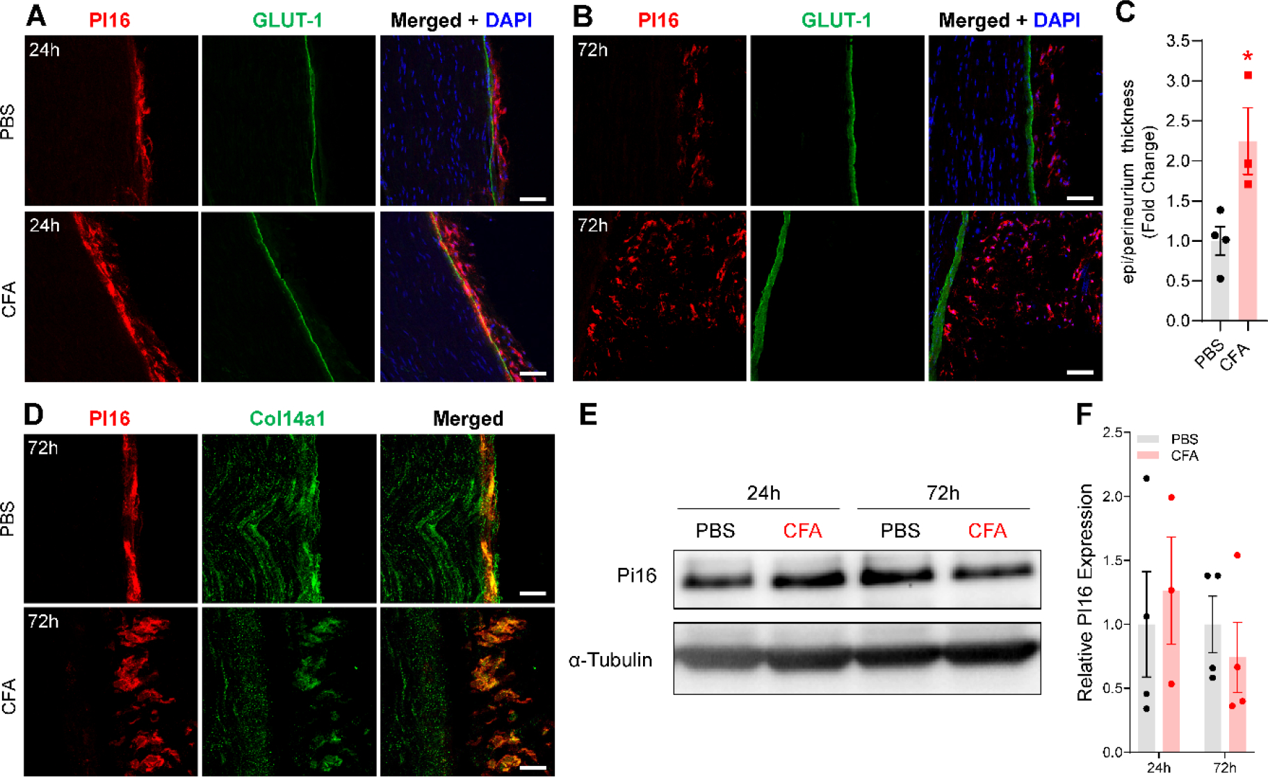
Fig. 3.

Effect of intraplantar CFA on PI16 protein expression. (A) Representative confocal image of sciatic nerve from PBS and CFA-treated female mice on day 1 of treatment showing immunostaining of PI16 (red) with outermost perineurial marker GLUT1 (green; B) from PBS and CFA-injected mice 3 days post-injection showing immunostaining of PI16 (red) with outermost perineurial marker GLUT1 (green). (C) Quantification of epineurium/perineurium thickness measured from perineurial marker GLUT1. Statistical analysis was performed using t-test: *P<0.05. (D) Representative image of PI16 (red) and Col14a1 (green) staining in the sciatic nerve on day 3 after CFA injection. Merged images show the co-localization of PI16 and Col14a1 (yellow), merged images in CFA show expansion of Col14a1 positive fibroblast in the epi/perineurium of the sciatic nerve co-expressing PI16. Immunofluorescence data are representative of n=4, each group (Scale bar 50 μm). (E) Representative Western blot and quantification of sciatic nerve PI16 24 or 72h after CFA injection in WT mice, quantified in (F).
