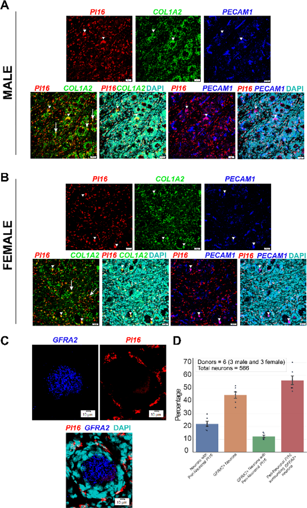
Fig. 7.

PI16 mRNA expression colocalizes with fibroblast marker COL1A2 in human DRG and surrounds GFRA2+ neurons. PI16 (red) expression overlaps with a subset of COL1A2+ fibroblasts (green) but not with endothelial cell marker PECAM1 (blue) in human DRGs recovered from male (A) and female (B) donors. Long arrows point to areas where PI16 mRNA puncta overlap with COL1A2 mRNA. White arrow heads show lipofuscin auto-fluorescence signal present in all channels. Scale bar = 50 μm. (C) PI16 mRNA (red) surrounds neurons, including GFRA2+ neurons (blue) in human DRG. (D) Quantification of PI16 mRNA puncta shows that approximately 20% of all human DRG neurons have peri-neuronal PI16 and about 10% of GFRA2+ neurons have peri-neuronal PI16. Scale bar = 10 μm.
