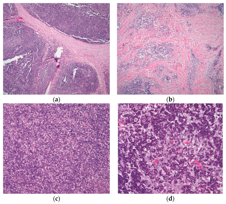
Figure 2.
The different growth patterns of conventional thymoma: (a) type B1 thymoma showing lobulation by thick fibrous bands; (b) thymoma in areas of fibrocollagenous stroma with no well-defined lobulation; (c) thymoma with an even distribution of lymphocytes and epithelial cells and a diffuse development pattern; and (d) higher magnification displaying the mixed population of lymphocytes and polygonal epithelial cells. (a,b) (H&E, 4×); (c) (H&E, 10×); (d) (H&E, 20×).

