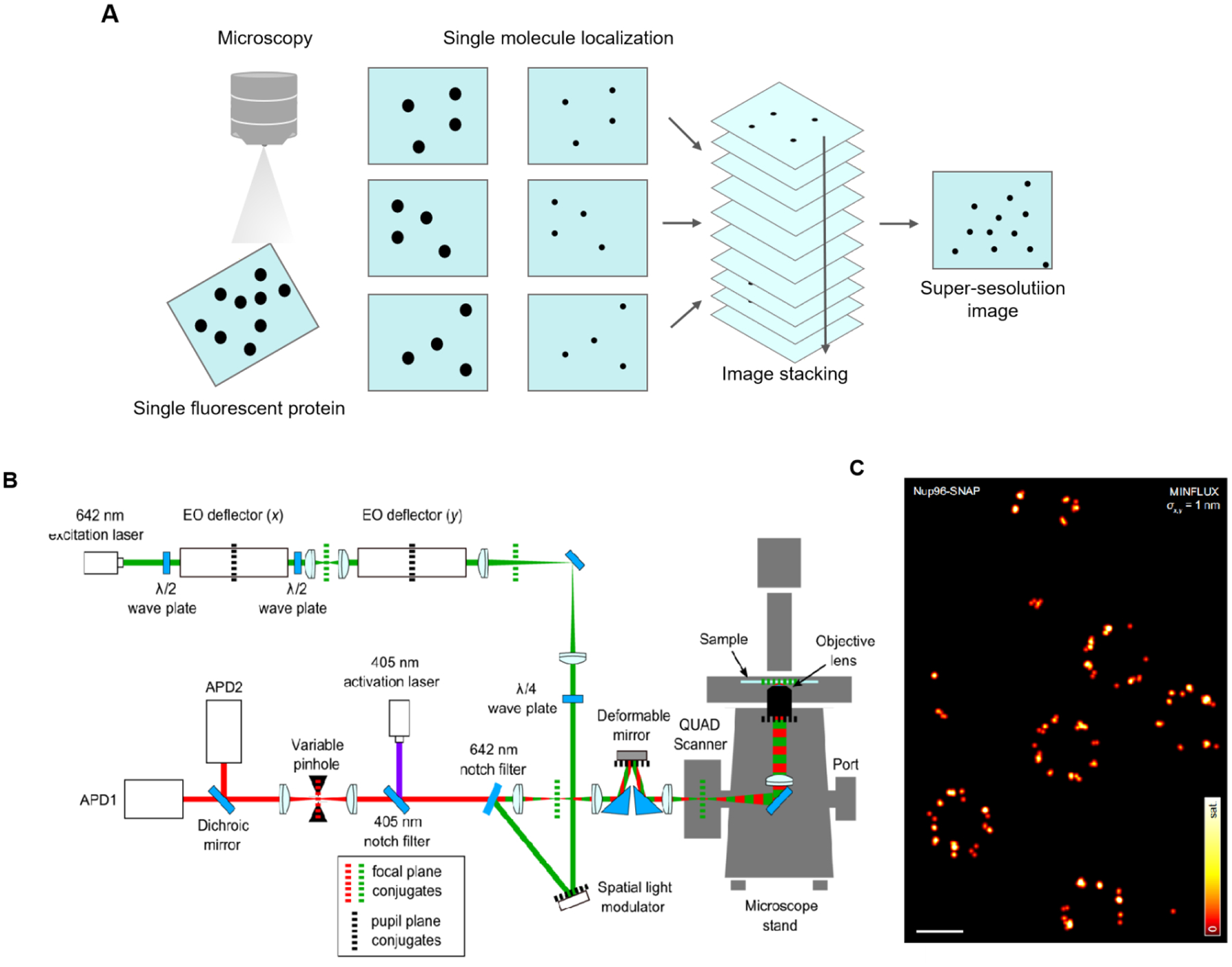
Figure 2.

Principle and application of SRM based on SMLM. (A) Principle of SMLM. (B) MINFLUX optical arrangement. An excitation beam (shown in green) is electro-optically deflected in x,y plane, spatially phase-modulated for conversion into a donut-shape, overlapped with an activation beam (purple), and after passing a deformable mirror and a galvanometer scanner in a 3D scanning assembly, it is focused into the sample on top of an all-purpose inverted microscope stand. Fluorescence from the sample (red) is descanned by the scanning assembly and passed to a variable confocal pinhole for detection using two avalanche photodiodes (APD). A stabilization unit based on both near-infrared scattering from fiducial markers and active-feedback correction provides sub-nanometer stability. (C) Principle of MINFLUX nanoscopy. MINFLUX fluorescence imaging of labeled cellular ultrastructure down to 1 nm (standard deviation) in fluorophore precision. σ: the precision obtained from a combined analysis of the statistical localization spread (standard deviation) in x and y. Fig. 2 B, C reproduced from Ref [70]. Scale bars: 200 nm.
