
Figure 8.
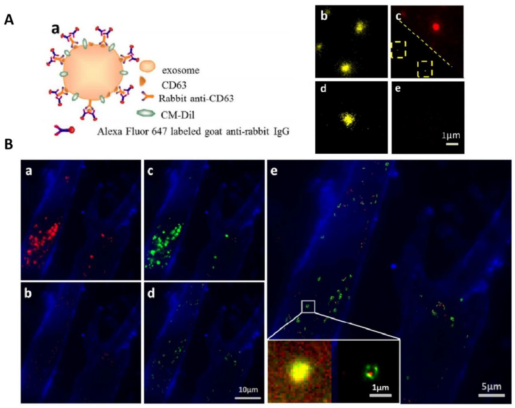
TIRF and PALM/STORM imaging of HeLa exosomes. (A) (a) Schematic illustration of indirect IF labeling of CD63 and exosome membrane stained with CM-Dil. (b) TIRF image of HeLa-derived exosomes stained with CM-Dil. (c) IF labeling of CD63 with Alexa Fluor 647, conventional diffractionlimited image (upper right) and PALM/STORM image (low left). (d) Immunofluorescence control sample without primary antibody for specificity detection, CM-Dil channel. (e) Immunofluorescence control sample without primary antibody for specificity detection, Alexa Fluor 647 channel. (B) Colocalization of SKBR3 exosomes and MRC-5 lysosomes. (a) TIRF image of internalized SKBR3 exosomes (red) and MRC-5 membrane (blue). (b) PALM/STORM image of the same region as shown in (a). (c) TIRF image of MRC-5 lysosomes (green) and MRC-5 membrane (blue). (d) PALM/STORM image of the same region as shown in (c). (e) Colocalization of MRC-5 lysosomes (green) and internalized SKBR3 exosomes (red). Fig. 8 reproduced from Refs [130].