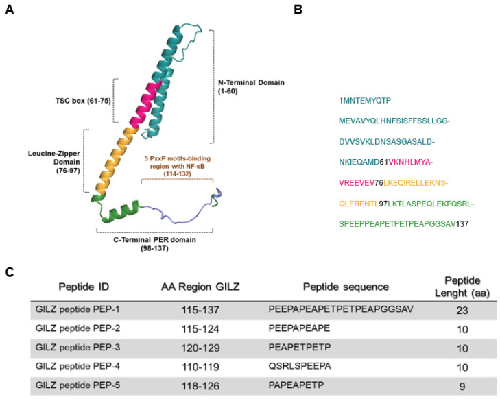
Figure 1.
Predictive structure of GILZ protein. (A) A three-dimensional representation, created by COSMIC2 platform (https://cosmic-cryoem.org, accessed on 15 September 2023) and modified with PyMOL Molecular Graphics System (http://www.pymol.org/pymol accessed on 15 September 2023), of GILZ protein constituted by four domains: a N-terminal domain (NTD, 1–60 aa), TSC-box (61–75 aa), a leucine zipper (LZ, 76–97 aa), and a PER region spanning from 98 to 137 aa residues. (B) The primary murine sequence of GILZ protein. (C) The table represents sequences of five GILZ peptides spanning different portions of the PER region, which is responsible for GILZ-NF-κB protein-to-protein interaction [21].

