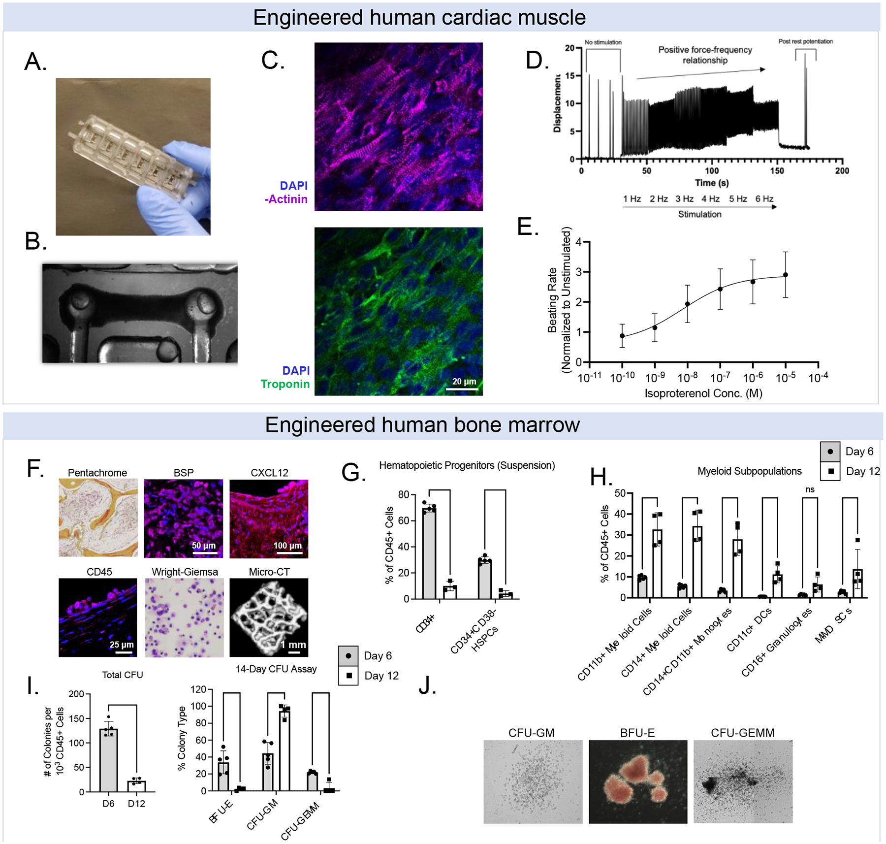
Figure 2. Baseline properties of engineered human tissues. Engineered cardiac muscle tissues (eCT).

(A) Bioreactor platforms. (B) Example bright field image of eCT on flexible pillars. (C) Example image of aligned cardiomyocytes stained for a-actinin (magenta), cardiac troponin (green), and DAPI (blue); scale = 20 μm. (D) Force-frequency relationship for eCTs. (E) Responses of eCTs to isoproterenol, a beta-adrenergic agonist. Engineered bone marrow tissues (eBM). (F) Histological staining of key hematopoietic and downstream blood/immune markers. (G) Characterization of CB-derived CD34+ cells and CD34+CD38− hematopoietic stem progenitor cells (HSPCs) in eBM tissues over 1–2 weeks. (H) HSPCs in human eBM begin to differentiate into myeloid cells (i.e., monocytes, dendritic cells, granulocytes) over 1–2 weeks. (I, J) Multipotency of HSPCs over 1–2 weeks using colony forming unit assays. Data are shown as mean +/− SD. *p<0.05, ** p<0.01, *** p<0.001, **** p<0.0001.
