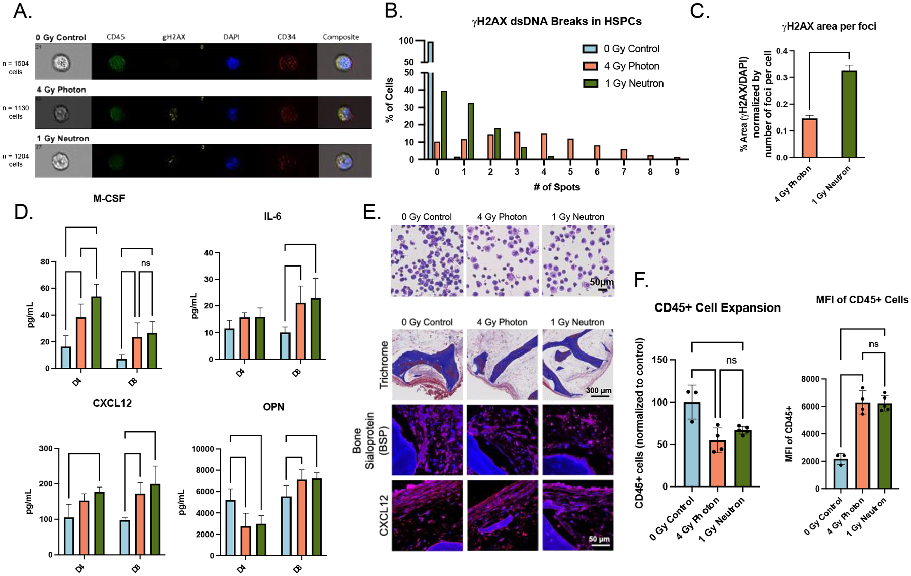
Figure 4. Ionizing radiation causes changes to engineered bone marrow (eBM).

(A) Representative images of DNA-damage marker γH2AX in hematopoietic cells isolated 1-hour post-radiation. (B-C) Quantitative analysis of the numbers and average area of dsDNA breaks (normalized to foci per cell) with unpaired t-test. (D) Acute doses of radiation caused early signs of inflammation (M-CSF, IL-6) and stromal damage (CXCL12, OPN) in supernatant. (E) Histological staining of eBMs 3-weeks post-radiation. (F) CD45+ cell proliferation and median fluorescent intensity 3-weeks post-radiation using flow cytometry. Data are shown as mean ± SD. *p<0.05, ** p<0.01, *** p<0.001, **** p<0.0001.
