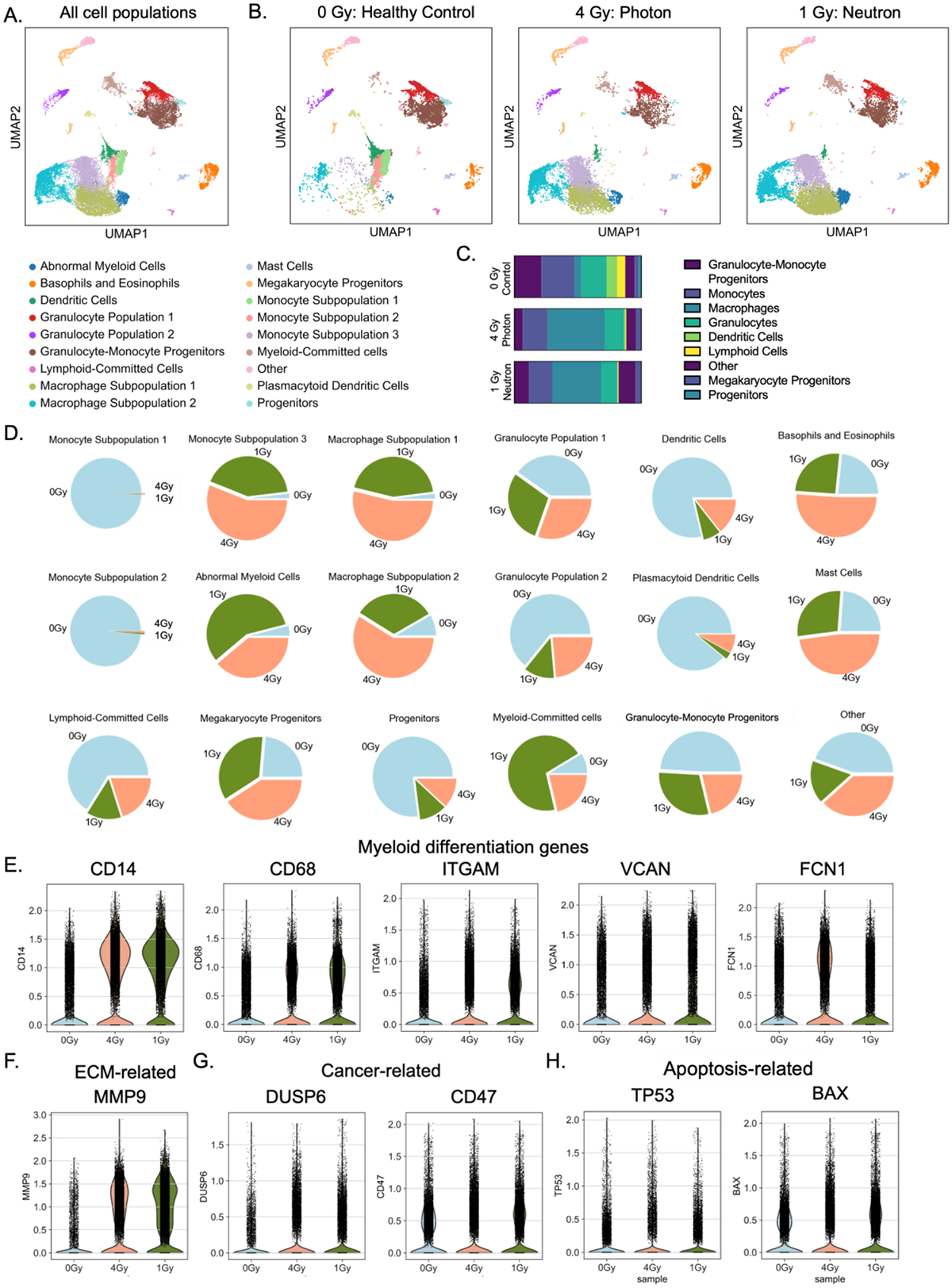
Figure 5. Single-cell transcriptomics reveals myeloid skewing in irradiated eBMs.

(A) Uniform Manifold Approximation and Projection (UMAP) dimensionality reduction to identify blood and immune cell progeny after 3 weeks of culture. (B) Breakdown of UMAP clusters for each experimental condition. (C) Distribution of the main classes of immune cells, showing increased myeloid population (mainly in macrophages) in irradiated groups. (D) Cell distributions for each experimental condition within the 18 clusters. (E-H) Differential expression analysis of neutron irradiated eBMs reveals increased matrix remodeling. Violin plots showed increased expression of genes related to (E) myeloid differentiation, (F) matrix degradation, (G) cancer, and (H) apoptosis.
