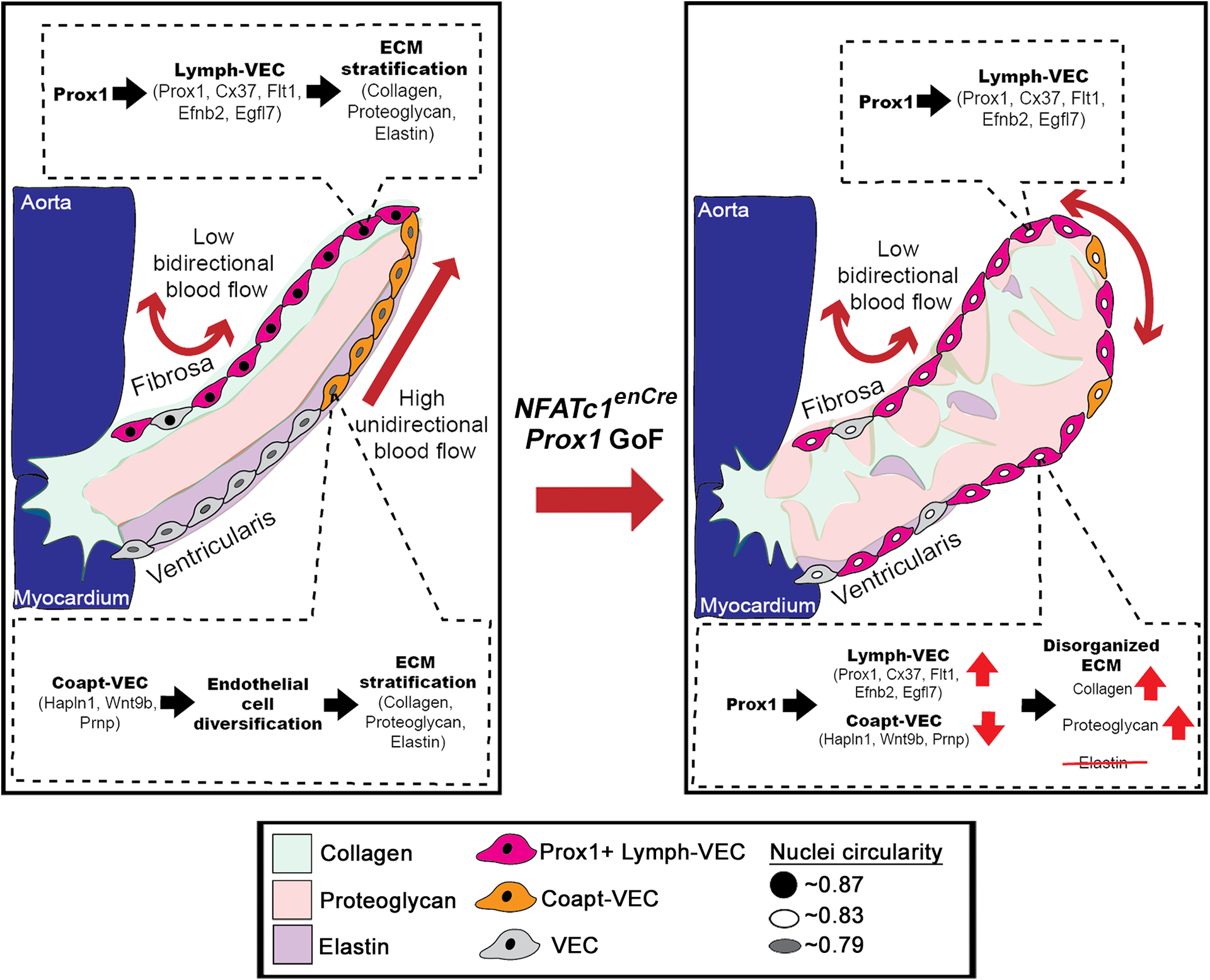
Figure 7. Model of Prox1 regulation of aortic valve endothelial specification compared to NFATc1enCre Prox1 GoF which disrupts endothelial diversity, leading to myxomatous extracellular matrix.

In a healthy valve, the Prox1+ Lymph-VEC population (pink cells) is localized to the fibrosa side of the AoV and is required for the development of the stratified trilaminar extracellular matrix (ECM) found in heart valves. The Coapt-VEC population (orange cells) are normally localized to tip of the leaflet on the ventricularis side of the AoV. Ectopic induction of Prox1 on the ventricularis side of the AoV increases Lymph-VEC (pink cells) and decreases Coapt-VEC (orange cells) gene expression on the ventricularis side of the AoV, disrupting VEC diversification and leading to a disorganized myxomatous ECM. Collagen (green). Proteoglycan (red), Elastin (purple).
