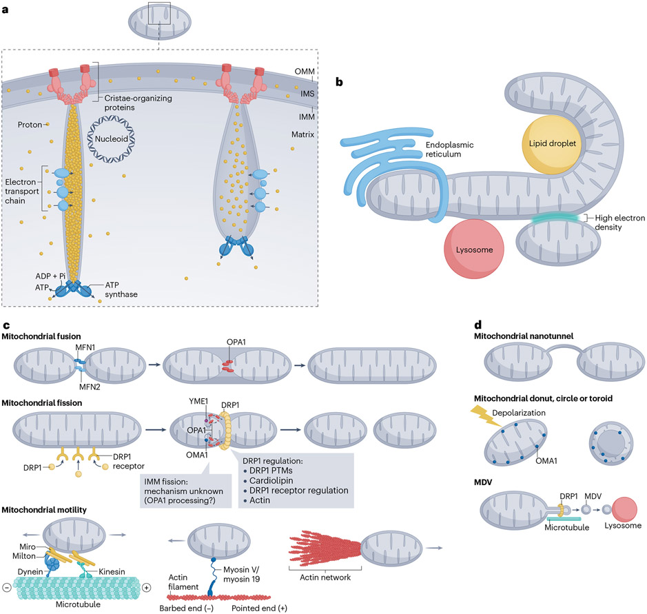
Fig. 2 ∣. Mitochondrial structure and dynamics.
a, Mitochondrial structure. The double-membrane structure (outer mitochondrial membrane (OMM) and inner mitochondrial membrane (IMM)) results in two aqueous compartments: the inter-membrane space (IMS) and the matrix. The IMM is segregated into the cristae membrane and inner-boundary membrane. Cristae junction proteins (light red) maintain a tight opening at the cristae base and interact with proteins mediating OMM-IMM tethering (dark red). The four complexes of the electron transport chain (light blue) enrich on the side of the cristae, whereas ATP synthase (dark blue) enriches at the cristae tip. A nucleoid, containing compacted mitochondrial DNA, might attach to the cristae side. Shown here are two cristae, one with a tight cristae junction and high proton gradient (left), and the other with a more open cristae junction and lower proton gradient (right). b, Mitochondrial contacts with the endoplasmic reticulum, lipid droplets, lysosomes and other mitochondria. c, Core components of mitochondrial fusion (MFN1, MFN2, OPA1), fission (DRP1, DRP1 receptors) and motility (microtubule based by kinesins or dynein, myosin based by myosin V or myosin 19, actin network based). Hypothetical mechanism of IMM fission, mediated through cleaved OPA1 (red) generated by either the YME1 (purple) or OMA1 (blue) protease are also shown. Dynactin complex for dynein motility is not shown for simplicity. Actin network for polymerization-based motility is not drawn with a specific architecture, which is undefined at this point. d, Other forms of mitochondrial dynamics. Top: nanotunnels between two mitochondria, with both OMM and IMM in a nanotunnel. Middle: mitochondrial IMM reorganization into toroids (also called donuts or circles), without OMM fission. This rearrangement occurs upon acute mitochondrial depolarization and is dependent on OMA1. Bottom: mitochondrial-derived vesicle (MDV) formation through membrane tube extrusion (microtubule-dependent)and DRP1-mediated constriction. PTMs, post-translational modifications.

