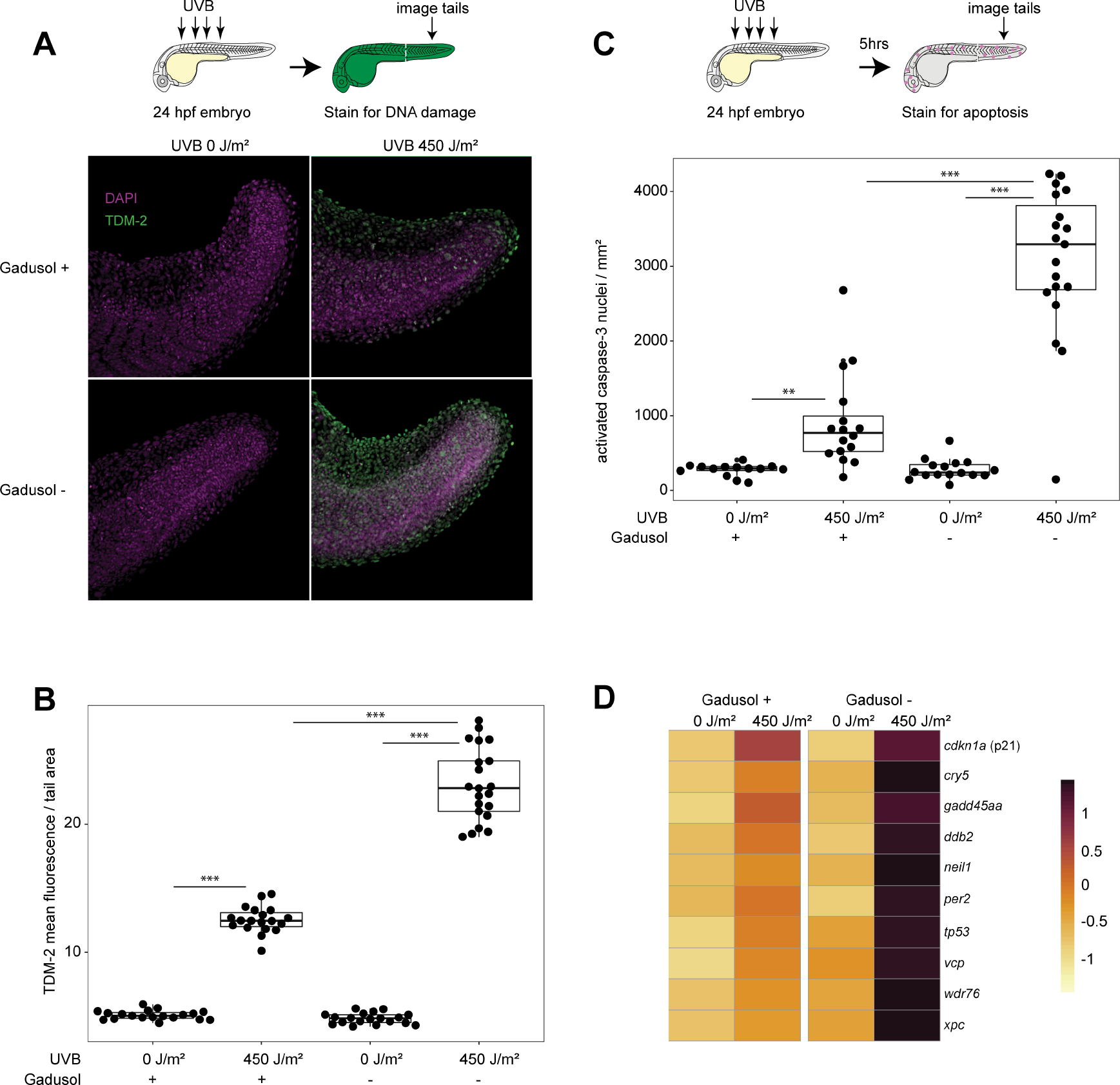
Figure 2. Gadusol functions as a sunscreen preventing DNA damage and apoptosis.

A, Immunohistochemistry, using an antibody that recognizes CPDs (TDM-2), on 24 hpf embryos immediately after mock or UVB exposure. Representative images shown.
B, Quantification of CPD labeling normalized to tail area (mm2). From left to right, n = 19, 19, 19, 21; N = 2 for all groups.
C, Quantification of immunohistochemistry, using an antibody that recognizes activated caspase-3. n = 14, 16, 16, 20. N = 2 for all groups.
D, Significant upregulation of select UVR response and DNA damage GO term-associated genes measured from the indicated conditions and genotypes using RNAseq on 24 hpf embryos after mock exposure or UVB exposure. RNA was collected 5 hours post mock or UVB exposure. Gene expression is scaled by rows. Significance determined via Fishenricher.26.
Student’s T-test P*<0.05; P**<0.01; P***<0.0001. n = number of embryos. N = number of clutches. See also Figure S3, Data S3, and Table S1.
