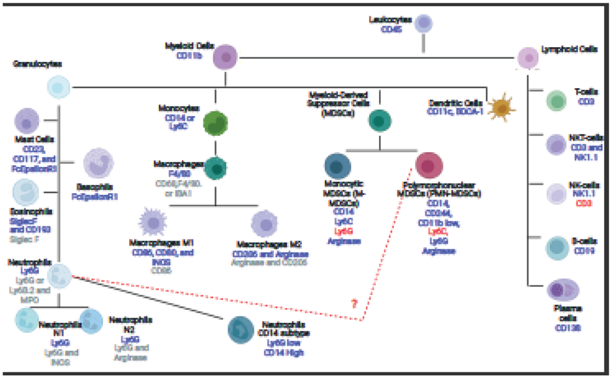
Fig. 1. Diagram of Identification Markers to Separate Neutrophils from Basophils, Eosinophils, Mast cells, Macrophages, and Monocytes.

The diagram shows the different cell types within the myeloid and lymphoid lineages. The blue text under each cell type indicates a positive flow marker for that cell type, while the red text indicates a cell marker that is not present in the cell type. The grey text for macrophages, neutrophils, and eosinophils are markers that can be used in immunohistofluorescence to detect these cell types. This list is not an extensive list of cell types as some can be further divided, nor an exhaustive list of cellular markers.
