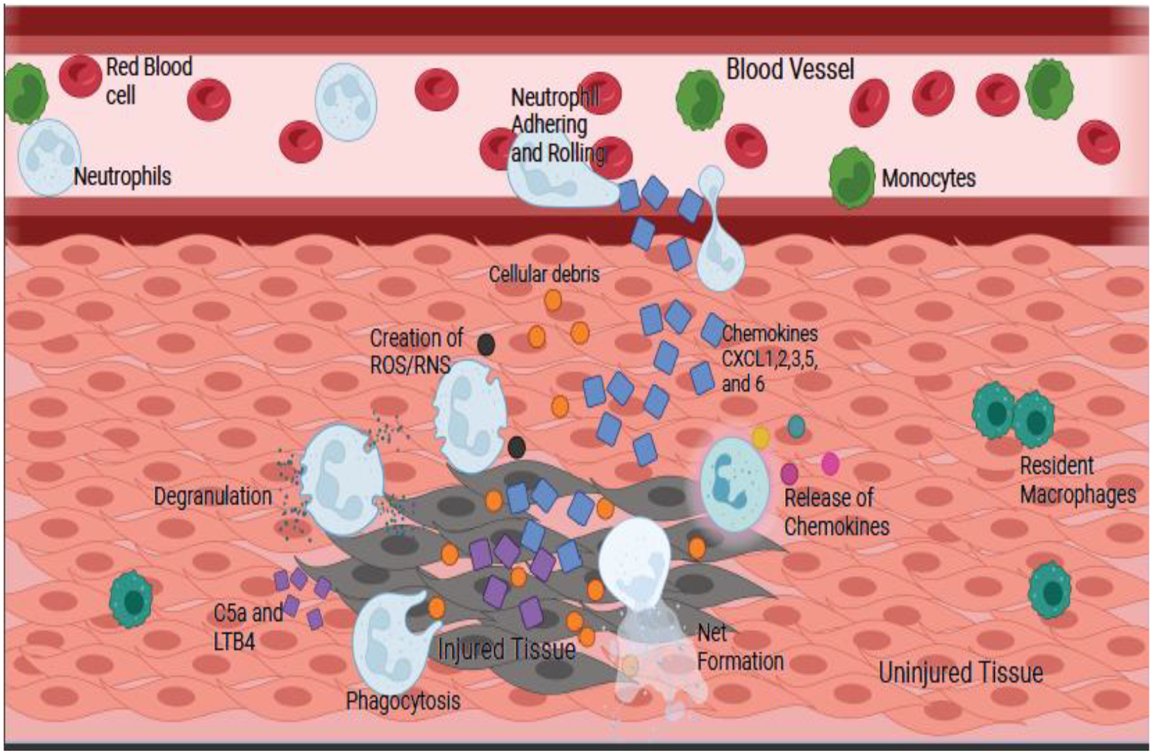
Fig. 3. Neutrophil Chemotaxis and Mechanism of Action.

Neutrophils will follow the gradient of chemokines to the injury site, then adhere and roll along the vessel before firmly adhering and entering the tissue. Entering the tissue is accomplished by degranulation of the tertiary granules. Once in the tissue, the cytokines CXCL1, 2, 3, 5, 6, LTB4 and C5a, which are released by the injured tissue, will trigger different cellular functions leading to degranulation, phagocytosis, creation of reactive oxygen and nitrogen species (ROS/RNS), formation of NETs, and release of chemokines. The orange circles are cellular debris, the blue squares are chemokines, and the purple squares are C5a and LTB4. The cells are labeled in the figure.
