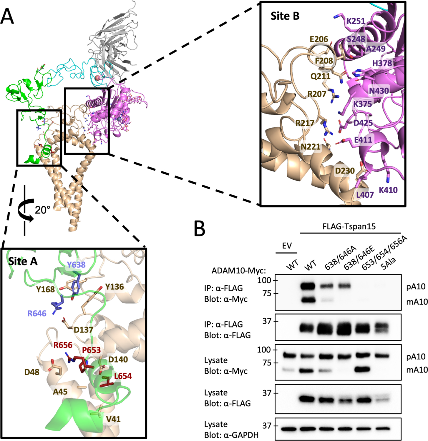
Figure 2. ADAM10-Tspan15 contact interfaces and mutational analysis.

(A) Cartoon representation of the vFab-ADAM10-Tspan15 complex with site A (left) and site B (right) interfaces boxed. A close-up view of the interface at site A is shown below the main panel, and a close-up view of the site B interface is shown to the right. The two clusters of interface A residues mutated in the immunoprecipitation assays are shown in blue and maroon. (B) Effect of ADAM10 mutations on co-immunoprecipitation with Tspan15. HEK293 cells were co-transfected with wild-type or mutant ADAM10-myc and FLAG-Tspan15 or vector control, and cell lysates were immunoprecipitated with anti-FLAG antibodies. The lysates and immunoprecipitates were subjected to SDS-PAGE, and analyzed by western blot using anti-myc or anti-FLAG antibodies. Lysates were also probed with an anti-GAPDH antibody (bottom) as a loading control. See also Figure S4.
