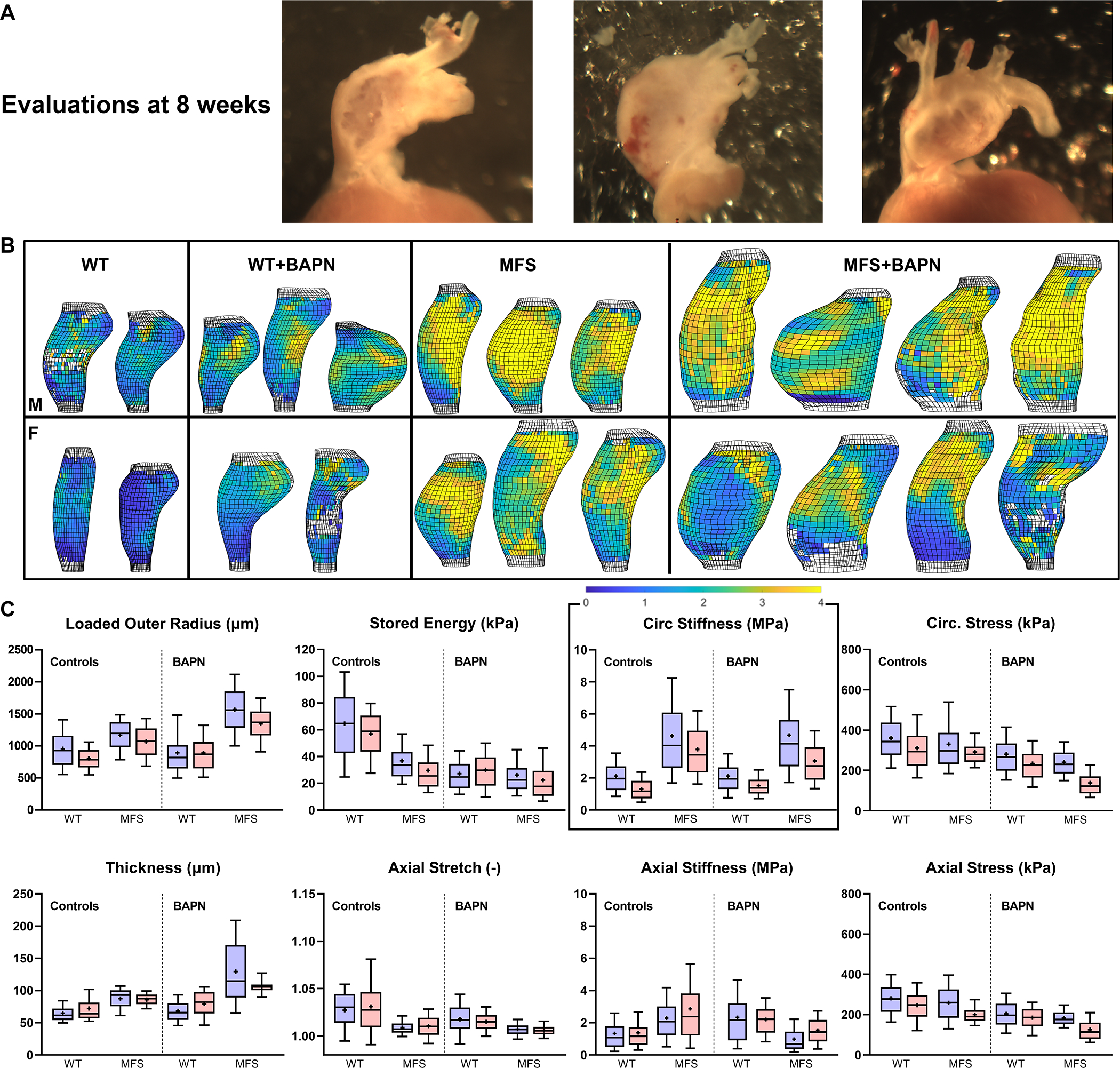
Figure 4.

A. Illustrative gross images of three male MFS aortas following 4 weeks of BAPN that started at 4 weeks of age (then evaluated at 8 weeks of age). Note the marked regional thinning of the wall even though these vessels did not rupture in vivo. B, C. Multimodal (pDIC + OCT) ex vivo testing under physiological loading confirmed findings from standard biaxial testing in a randomly selected sub-set of 8-week old male (M) and female (F) mice ( of 40), but further revealed regional heterogeneities in most geometric and mechanical metrics (see Figs S9–S12 for sample-specific distributions). Highlighted in panel B, for illustrative purposes, is the perhaps surprising lack of a dramatic effect of in vivo BAPN on circumferential material stiffness – with a colorimetric scale bar given from ~0 (darkest blue) to > 4 (yellow) MPa. Each bar and whisker plot represents ~2000 to 4000 data points, that is, ~1000 results around the circumference and along the length of each specimen, thus ~23000 data points are represented and compared within each of these 8 panels.
