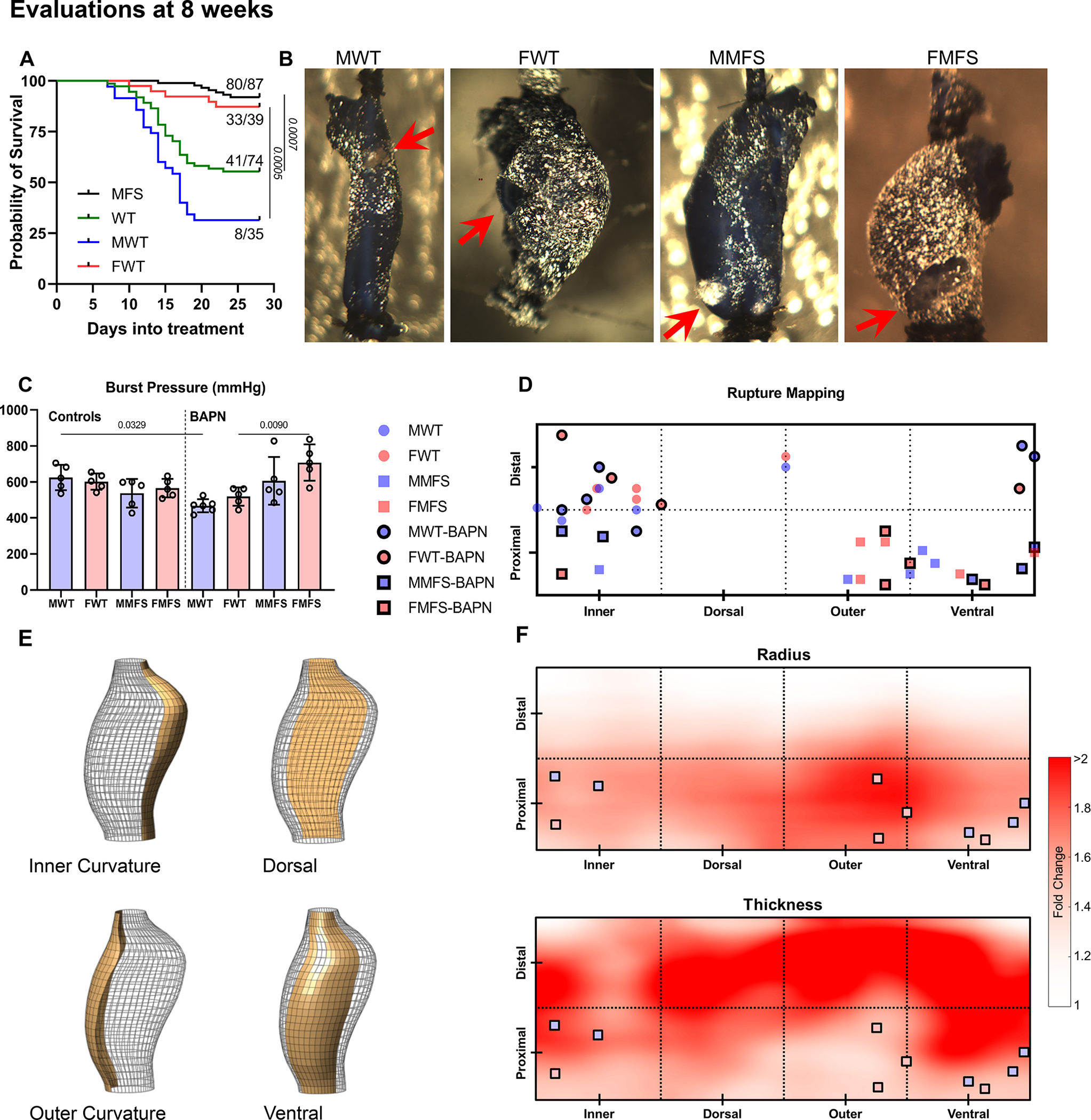
Figure 5.

A. Survival curves reveal a statistically higher BAPN-associated lethality in young WT than in young MFS mice, particularly in males (27/35 male WT mice died, 2/42 male MFS mice died). All recorded deaths were natural and mice were not necropsied to determine cause of death. B, C. Albeit restricted to aortas from surviving mice, ex vivo burst-pressure testing (mean±SD) confirmed greatest vulnerability in the young male WT aortas (red arrows indicate rupture sites). D, E. Rupture sites mapped onto four circumferential (inner curvature, dorsal, outer curvature, and ventral) and two axial (proximal half vs. distal half) regions. F. Maps show spatial fold-changes of radius and thickness associated with MFS and BAPN relative to the non-exposed WT controls (see Figure S13 for separate effects of sex, genotype, and BAPN). Dilatation in the MFS-BAPN group was observed primarily in proximal regions, particularly near the outer curvature, while thickening was mostly observed distally. Locations of failure during ex vivo testing of MFS aortas tended to coincide with regions that experienced local dilatation (i.e., increased radius) without substantial thickening.
