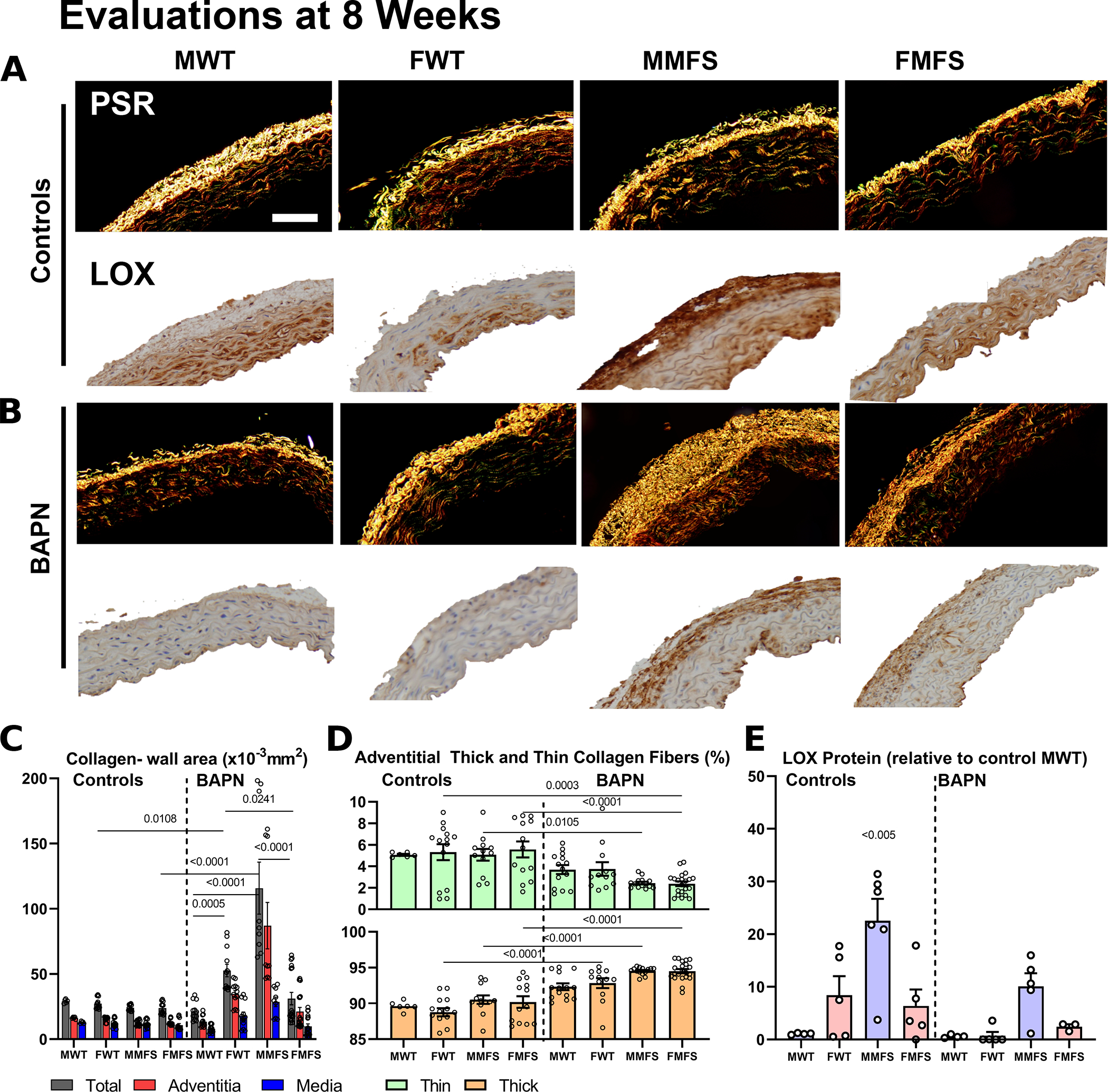
Figure 6.

A-D. Picro-sirius red stained cross-sections suggested modest changes in mural collagen following BAPN in older mice (not shown) as well as in most younger mice with the stark exception of young male MFS mice in which there was a marked increase in adventitial collagen. BAPN increased the ratio of thick:thin adventitial collagen in all young mice, perhaps due to a reduced incorporation of thin fibers during turnover. Scale bar = 100 μm. E. Notably, immuno-staining for LOX revealed generally higher levels in female than male mice, and higher levels in MFS than WT mice prior to BAPN. These levels tended to drop following BAPN although group-to-group trends persisted. Data are presented as means±SEM; statistical comparisons by non-parametric Kruskal Wallis test followed by Dunn’s post-hoc test for multiple comparisons; noting that all statistical comparisons with the controls MMFS group were found significant (noting on the graph as <0.005).
