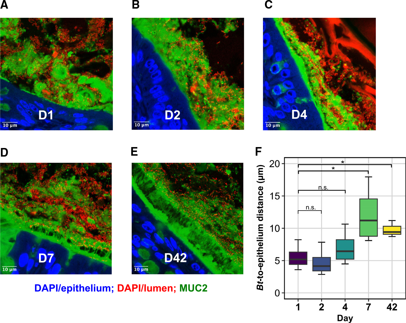
Figure 6. Bt localization shifts from the mucus toward the luminal space over colonization.

(A–E) Representative cross-sections of distal mouse colon fixed in Carnoy’s solution at (A) day 1 (D1), (B) D2, (C) D4, (D) D7, and (E) D42 of colonization by Bt. Blue, DAPI staining in the epithelium; red, DAPI staining in the lumen of gut, including bacteria, debris, and shed host nuclei; green, antibody staining for MUC2. Scale bars, 10 μm.
(F) Mean Bt epithelial proximity. n = 3–4/group, t test, *p < 0.05.
