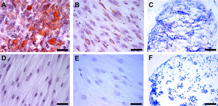
Figure 2.
In vitro differentiation potential of human Conj-MSCs. Cells differentiated into three different lineages. (A) Conj-MSCs differentiated into adipocytes. Lipids are marked in red by Oil Red staining. (B) Cells differentiated into osteocytes that have calcium deposits as seen in brown by Von Kossa staining. (C) Cells differentiated into chondroblasts, as observed in chondroblast pellet sections stained with Toluidine Blue dye that stain the proteoglycans in purple. Cells cultured in the same conditions but without differentiation media were also stained with Oil Red (D), Von Kossa (E), and Toluidine blue (F), but no lipids, calcium deposits, or proteoglycans were detected. Bar = 50 µm.

