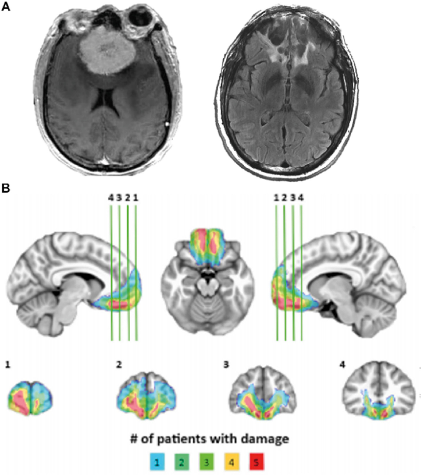
Fig. 1 –

VMPFC lesions. (A) Example lesion from one patient showing appearance of skull-base meningioma on preoperative clinical T1-weighted MRI (left) and cavitary parenchymal VMPFC lesion postoperative clinical T2-weighted MRI (right). (B) Lesion overlap map depicting lesion extent across the VMPFC lesion group. All subjects have bilateral lesions involving the ventral third of the medial frontal cortex and medial third of the orbitofrontal cortex. Colors indicate number of patients with damage in a particular region.
