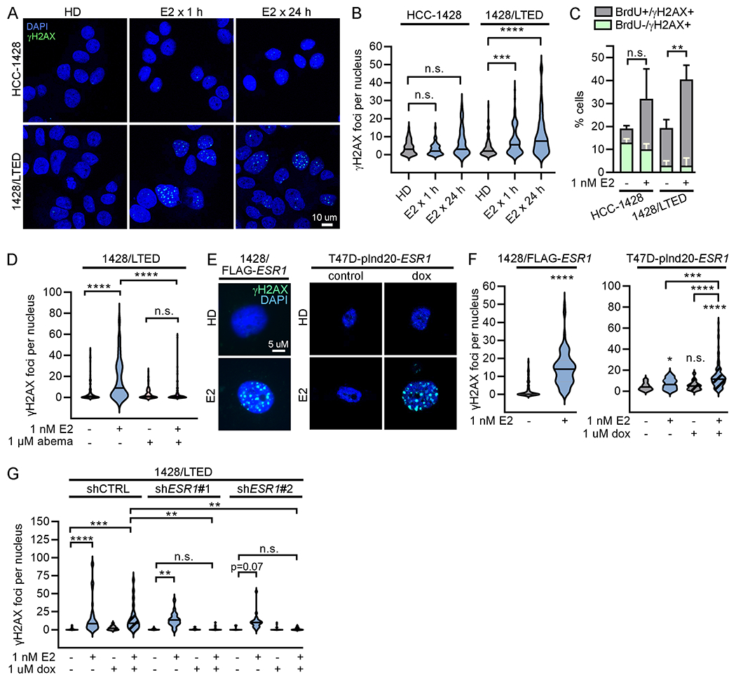
Figure 2. 17β-estradiol-induced DNA damage is dependent upon overexpression of ER.

(A/B) HCC-1428 (HD x 3 d) and 1428/LTED cells were treated ± 1 nM E2. Cells were fixed and stained for γH2AX (green) and DAPI (blue). γH2AX foci were counted in ≥100 nuclei/group. (C) HCC-1428 (HD x 3 d) and 1428/LTED cells were treated ± E2 for 21 h, and labeled with BrdU for another 3 h. Cells were stained for γH2AX, BrdU, and cleaved PARP for flow cytometry analysis. Cleaved PARP-positive cells were excluded from analysis. Proportions of cells with DNA breaks (γH2AX-positive) that were or were not in S-phase (i.e., did or did not incorporate BrdU) were plotted. Proportions of γH2AX+/BrdU+ cells were statistically compared. Data are shown as mean of triplicates + SD. (D) Cells were treated ± E2 ± abemaciclib x 24 h and analyzed as in (B). (E/F) T47D/pInd20-ESR1 cells were pretreated with HD x 7 d, and then treated with HD ± dox x 14 d prior to seeding. All cells were then treated as indicated x 24 h and analyzed as in (B). (G) 1428/LTED cell lines expressing dox-inducible shRNA targeting ESR1 (two independent constructs) or non-silencing control were treated ± dox for 2 d, and then treated ± E2 x 24 h and analyzed as in (B). In (A/E), representative images are shown. *p<0.05, **p<0.005, ***p<0.0005, ****p<0.0001, n.s. = not significant.
