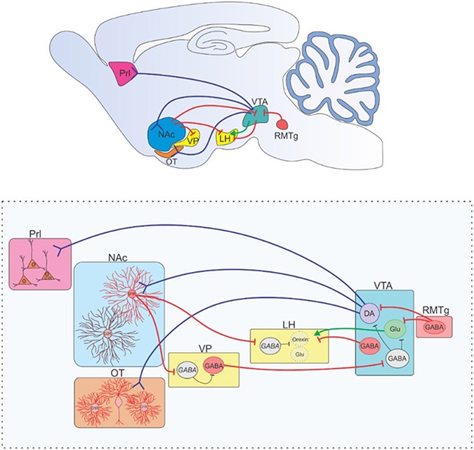
Figure 2.
Dopaminergic circuits involved in anesthetic emergence. (Top) A mid-sagittal view of a rodent brain is depicted with dopaminergic (blue), GABAergic (red), and glutamatergic (green) projections depicted summarizing the dopamine signaling pathways involved in regulating anesthetic emergence and detailed in the bottom panel. Activation of dopaminergic neurons in the VTA induce arousal under general anesthesia (33). Activation of the RMTg, a structure with dense inhibitory projections to VTA dopamine and glutamate neurons, enhances general anesthesia (60). Dopamine release from the VTA onto D1R-expressing neurons in the NAc (40, 42, 43) and OT (48) (Mesolimbic pathway) or the Prl (mesocortical pathway)(49) has been shown to induce emergence from general anesthesia in rodent studies. Recently, D1R NAc neurons projecting to the VP and LH have been shown to both induce emergence in rodents under general anesthesia(44). The target in the VP and LH are thought to be inhibitory interneurons. GABAergic VP projections have been shown to disinhibit VTA neurons, increasing arousal(45). VTA GABAergic neurons projection to the LH has been shown to induce emergence following anesthesia(61), and glutamatergic VTA projections to the LH enhance arousal(46). VTA = ventral tegmental nucleus, RMTg = rostromedial tegmental nucleus, NAc = nucleus accumbens, Prl = prelimbic cortex, VP = ventral pallidum, LH = lateral hypothalamus, OT = olfactory tubercle. Blue projections are dopaminergic, red are GABAergic, green are glutamatergic. Inhibitory interneurons are in black.

