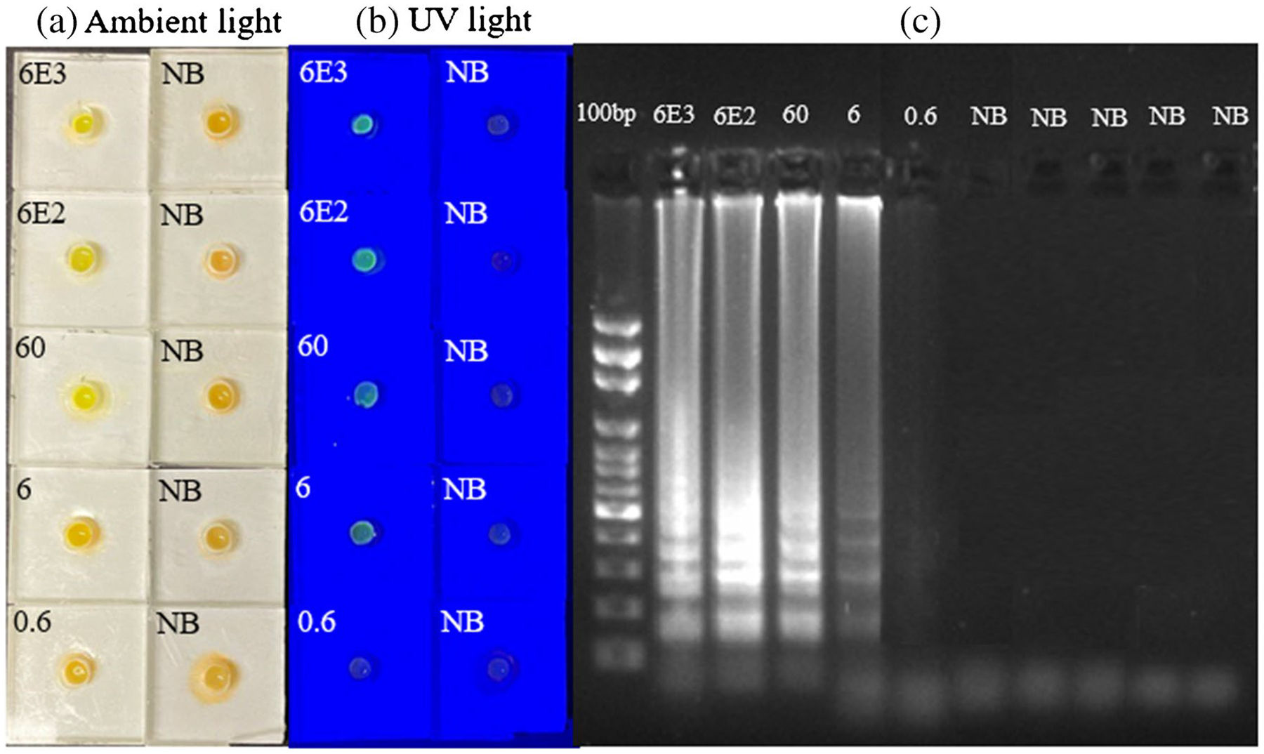
Fig. 4.

Endpoint detection of MAYV in whole blood samples using SPD. a. Pictures of the detection units under ambient light after RT-LAMP assay of whole blood samples spiked with cultured MAYV (viable virus) and blood samples without MAYV as negative controls (NB). b. Pictures of the same detection units in (a), illuminated by a blue LED flashlight. c. Gel electrophoresis image of the amplicons from each device in (a). The first lane is a DNA ladder while other lanes are for samples in (a)
