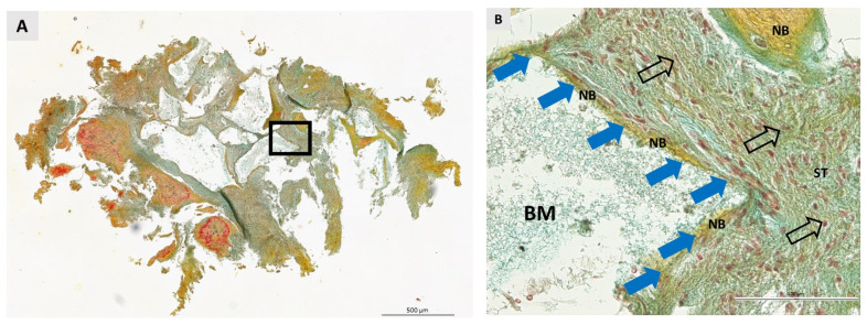
Figure 7.
Bone biopsy taken six months after ridge preservation with BXand stained with Movat pentachrome stain. (A) Longitudinal section. The square shows the area under higher magnification. Scale bar 500 μm. (B) The image shows an apposition line (blue arrows) in direct contact with the remaining biomaterial (BM). The soft tissue (ST) is rich in fibroblasts, whose nuclei are indicated by nonfilled black arrows. No signs of inflammatory reaction were detected in the specimens. Scale bar 100 μm.

