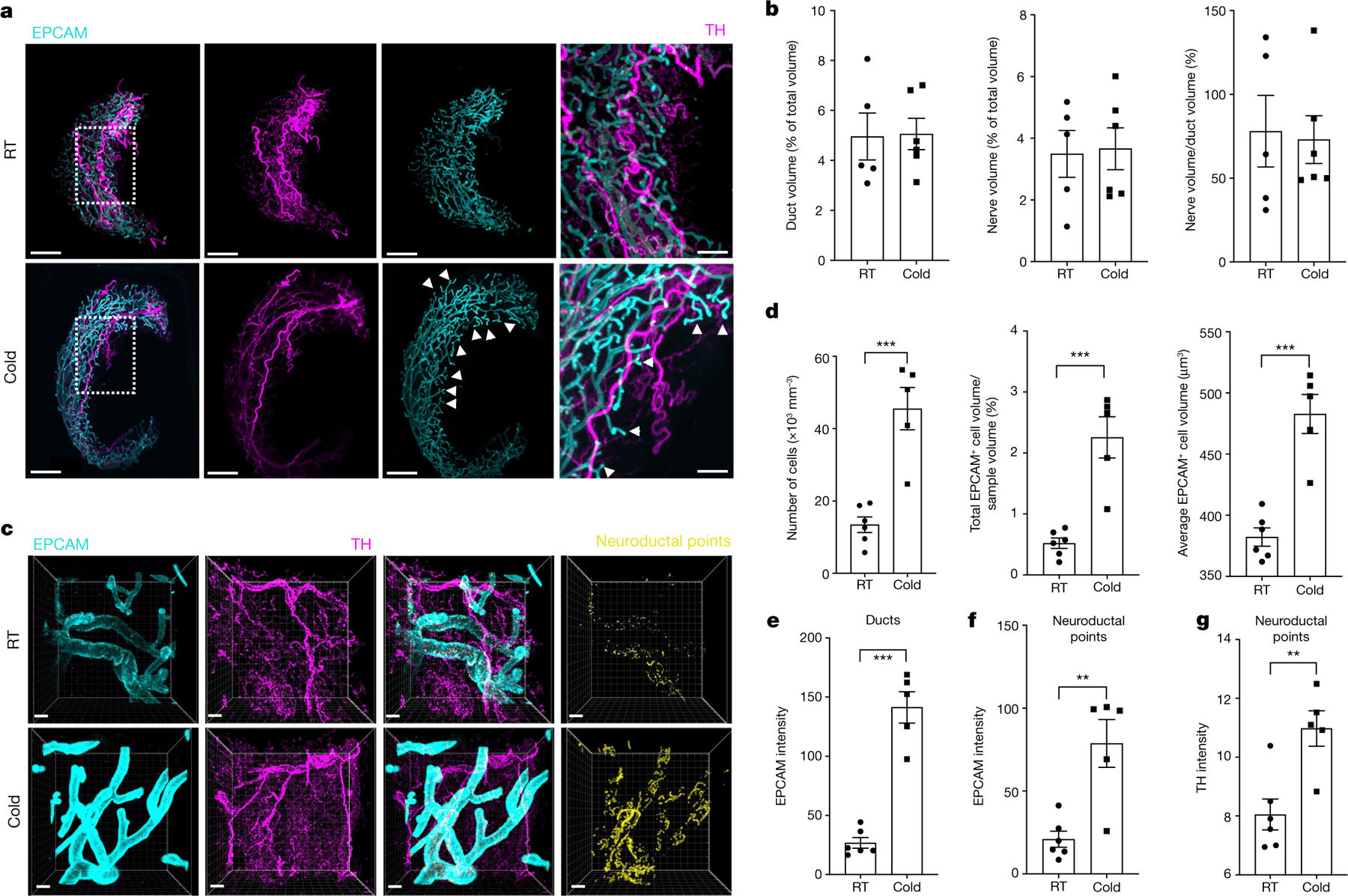
Fig. 2 |. SNS fibres directly innervate mammary ductal epithelium.

a, LSFM images of mgWAT isolated from female mice exposed to RT or cold for 24 h and stained with TH antibody (to mark SNS fibres) and EPCAM antibody (for ductal cells). Representative mgWAT images from six mice per condition. White arrows show terminal ductal bifurcations under the cold condition. Scale bars, 200 μm. b, Quantification of ductal volume and nerve volume as a percentage of total volume, and the ratio of nerve volume to ductal volume in RT or cold-exposed mgWAT. RT: n = 6; cold: n = 5. c, Confocal images of mgWAT isolated from female mice exposed to RT or cold for 24 h and stained with EPCAM and TH antibodies. Merged staining of EPCAM and TH represent neuroductal points. Representative image of six mice per condition. Scale bars, 100 μm. d–g, Quantification of EPCAM+ cells and cell volume (d), EPCAM intensity in ducts (e), EPCAM intensity at neuroductal points (f) and TH intensity at neuroductal points (g). RT: n = 6; cold: n = 5. b,d–g, Unpaired Student’s t-test. *P < 0.05; **P < 0.01; ***P < 0.001. Data are mean ± s.e.m.
