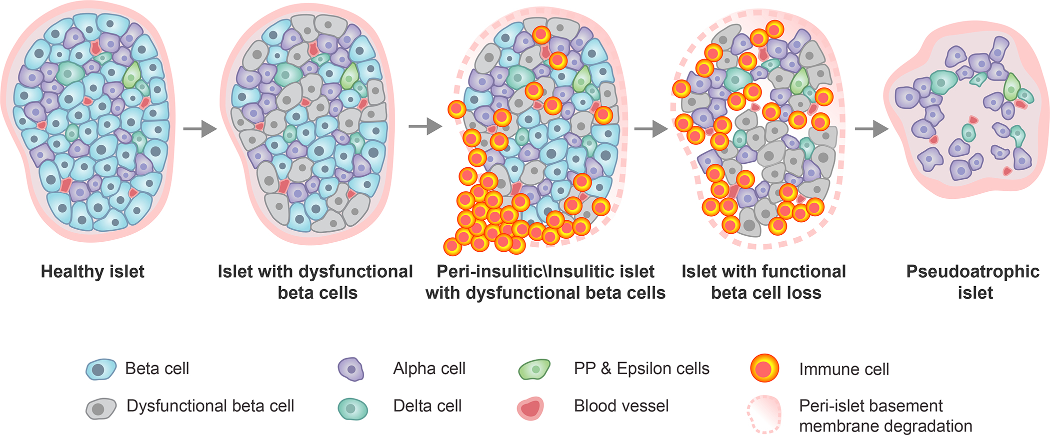
Figure 2. Histopathologic progression of type 1 diabetes within the human pancreatic islet.

Within an initially healthy islet, a portion of β-cells develop phenotypic and functional impairments. This precipitates immune cell infiltration around (peri-insulitic) and within (insulitic) the islet, coupled with degradation of the peri-islet basement membrane. Following the immune-mediated destruction of functional β-cells, the immune cells exit leaving behind an irregularly shaped, insulin-negative, pseudoatrophic islet characterized by presence of the remaining endocrine cell types (alpha, delta, PP, epsilon), reduced vessel diameter, increased microvascular density, and restored peri-islet basement membrane integrity. PP, pancreatic polypeptide.
