Fig. 1.

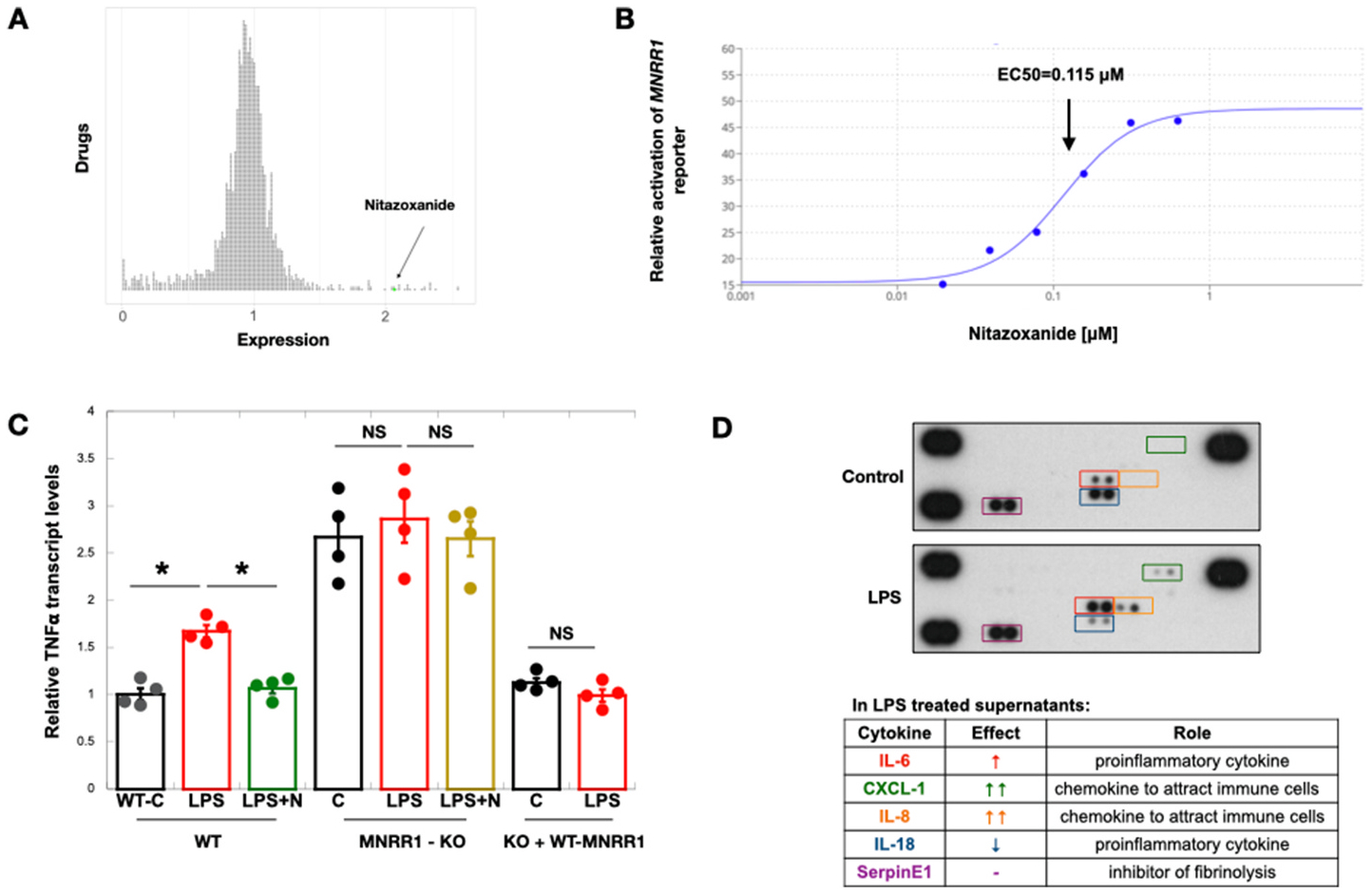
Nitazoxanide’s anti-inflammatory effects are via transcriptional activation of MNRR1.
A: Results of a screen of ~2400 FDA-approved drugs and natural compounds identified to transcriptionally activate (>1), inhibit (<1), or not affect (=1) MNRR1. Each circle represents one drug and the MNRR1 activator (nitazoxanide (N)) has been highlighted in green.
B: Equal numbers of HTR cells overexpressing the MNRR1-luciferase reporter were plated on a 96 well plate and treated with increasing amounts of nitazoxanide or vehicle (DMSO) for 24 h. EC50 was calculated using the activation of MNRR1 reporter relative to vehicle treated cells using the Quest Graph™ EC50 Calculator.
C: TNF transcript levels were measured in human placental cells. Actin was used as a housekeeping gene for normalization. Abbreviations: WT, MNRR1-WT; MNRR1-KO, knockout of MNRR1; C, control (water); LPS, lipopolysaccharide; N, nitazoxanide; KO + WT-MNRR1, knockout cells + MNRR1-WT overexpression. n = 4 biological replicates; significance: *, p-value<0.05; ns is non-significant.
D: Cell culture supernatants from control (water) or LPS-treated cells were tested as described in Materials and Methods for multiple cytokines using a membrane array. The sample in the bottom right corner is a blank negative control and the remaining
