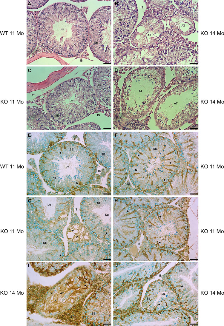
Fig 1.
LM of seminiferous epithelium (SE) of the testis stained with H&E (A-D) and anti-prosaposin antibody (E-J) of WT (A, E) and KO (B-D, F-J) mice at 11 or 14 months of age. In (F), several KO tubules demonstrate a normal appearance (NT) as seen in WT (A), while other KO tubules appear abnormal (AT) with a disrupted epithelium (B, D, G and I); some KO tubules only reveal Sertoli cells (B, G). Sertoli cells (arrowheads) immunolabeled for prosaposin are evident in WT (E) and KO (F-J) mice, as well as in the grossly altered tubules (G, I). Lu, lumen; IS, interstitial space. Scale bars = 35 μm.

