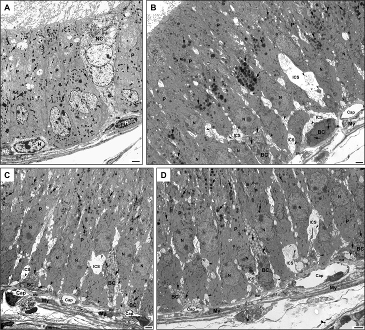
Fig 5.
EM of the initial segment of WT (A) and KO (B-D) mice. In (A), tall columnar principal cells (P) reveal a few small dense lysosomes (arrows). A halo cell (H) is adjacent to a small nondescript basal cell (BC), and a large narrow cell (NC) is noted. In KO mice, the dense lysosomes (arrows) appear to be more abundant and larger in size (B-D) as compared to WT mice (A). In (B-D), basal cells of KO mice reveal different shapes, sizes and commitment to the basement membrane and are filled with small to medium-sized pale lysosomes (arrows). Basally located dilated intercellular spaces (ICS) of KO mice contain membranous and vesicular profiles (arrowheads) (B-D). Cap, capillaries; My, myoid cells; BM, basement membrane; N, nucleus. Scale bars = 2 μm.

