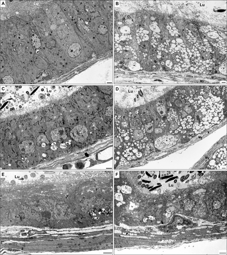
Fig 6.
EM of caput (A, B), corpus (C, D) and cauda (E, F) regions of WT (A, C, E) and KO (B, D, F) mice. In WT mice (A, C, E), principal cells (P) reveal few small dense supranuclear lysosomes (arrows) and occasional irregular larger dense lysosomes (Ly) infranuclearly (C, E). In all 3 regions of KO mice, principal cells (P) exhibit a plethora of medium to large-sized pale lysosomes (Ly) in their supra-and infranuclear areas (B, D, F), Clear cells (CL) are evident in KO mice (B, D, F) and at times show numerous pale stained lysosomes (F). Basal cells (BC) of KO mice (B, D, F) show prominent small to moderate pale stained lysosomes, which are not noted in WT mice (A). Large basally located eMPs (asterisks) reside in KO mice at the base of the epithelium (B, D). My, myoid cells; Lu, lumen; N, nucleus; H, halo cell. Scale bars = 2 μm.

