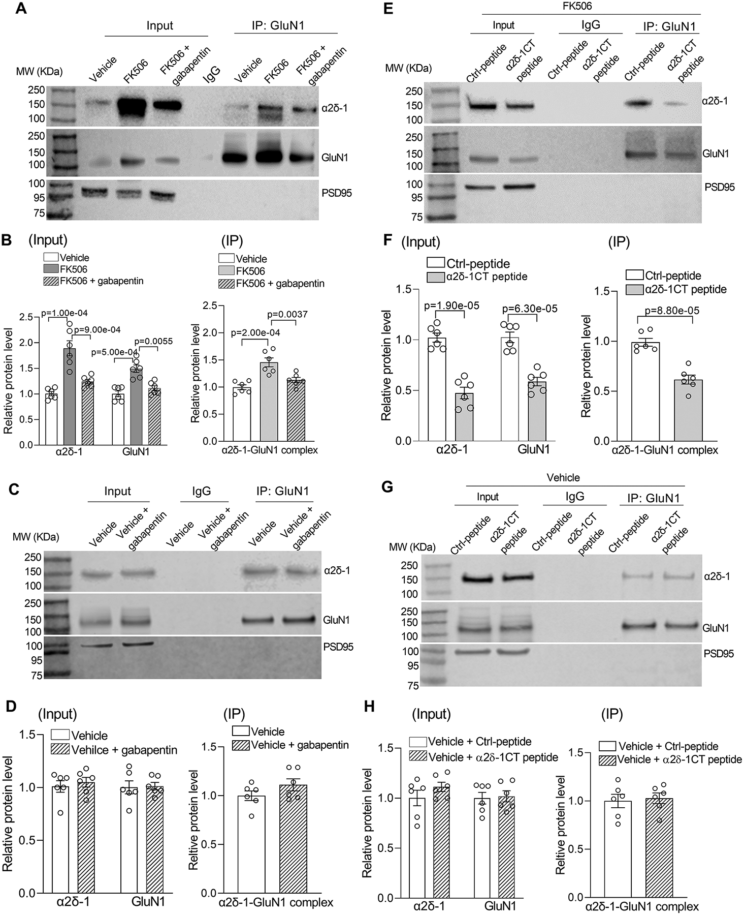
Figure 1. Calcineurin inhibition increases α2δ−1–NMDAR interactions and synaptic levels of NMDARs in the PVN.

A and B, Representative gel images (A) and qualification (B) show the effects of FK506 treatment with and without 100 μM gabapentin on protein levels of α2δ−1, GluN1, and α2δ−1–GuN1 complexes in PVN synaptosomes (n = 6 samples per group; each sample included PVN tissues from 2 male rats). C and D, Original gel images (C) and qualification (D) show the lack of effect of 100 μM gabapentin on protein levels of α2δ−1, GluN1, or α2δ−1–GluN1 complexes in PVN synaptosomes from vehicle control rats (n = 6 samples per group; each sample included PVN tissues from 2 male rats). E and F, Representative gel images (E) and qualification (F) show effects of pretreatment with 1 μM control peptide (Ctrl-peptide) or 1 μM α2δ−1 C terminus peptide (α2δ−1CT peptide) on protein levels of α2δ−1, GluN1, and α2δ−1–GluN1 complexes in PVN synaptosomes from FK506-treated rats (n = 6 samples per group; each sample included PVN tissues from 2 male rats). G and H, Original gel images (G) and qualification (H) show the lack of effect of α2δ−1CT peptide on protein levels of α2δ−1, GluN1, or α2δ−1–GluN1 complexes in PVN synaptosomes from vehicle control rats (n = 6 samples per group; each sample included PVN tissues from 2 male rats). MW, molecular weight. PSD95, a synaptic protein marker, was used as a loading control. One-way ANOVA with Bonferroni’s post hoc test was used in B; two-tailed Student’s t test was used in D, F, and H.
