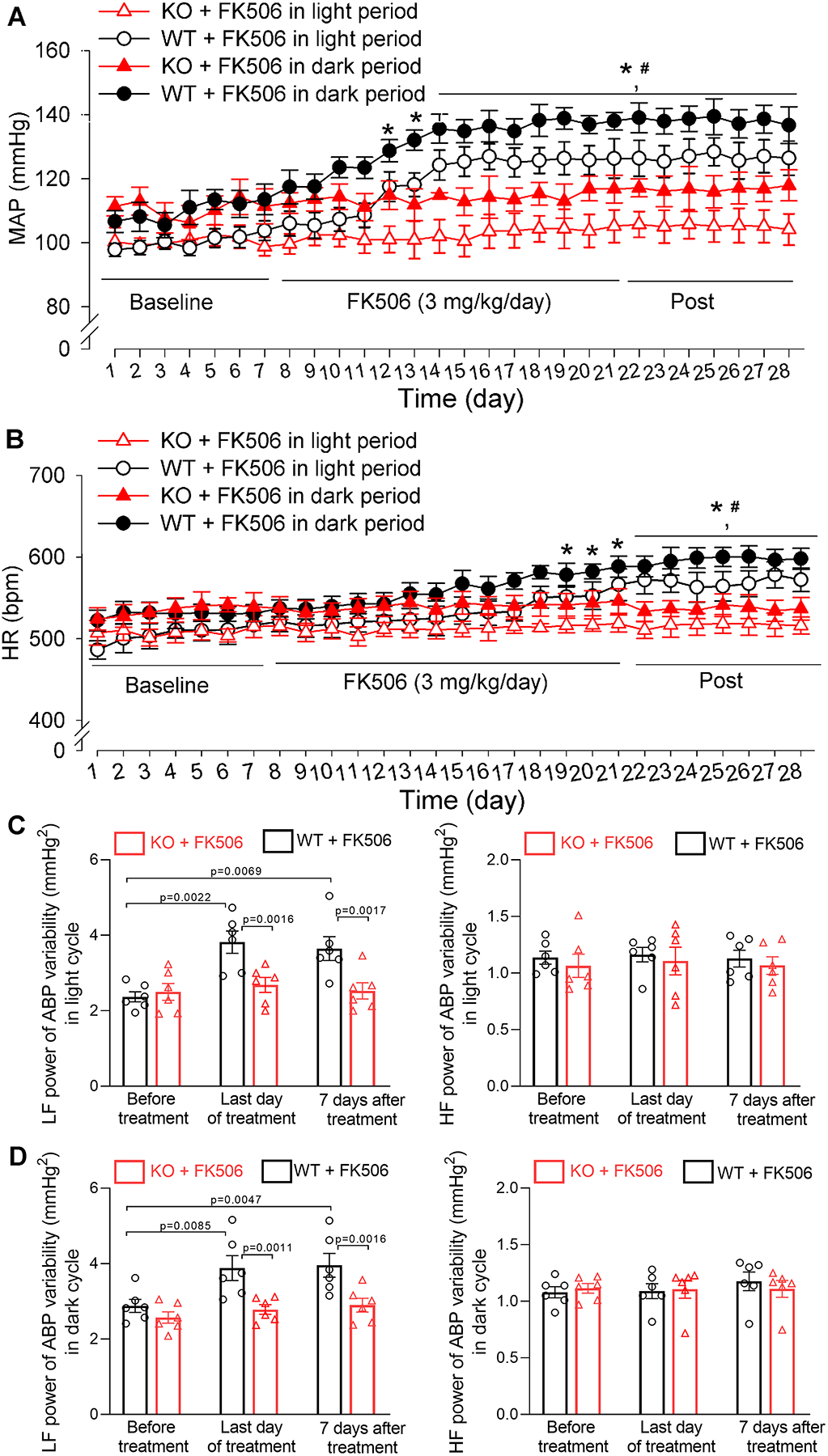
Figure 7. Genetic ablation of α2δ−1 prevents the development of CIH in mice.

A and B, Radiotelemetry recording data show the time course of changes in mean arterial blood pressure (MAP, A) and heart rate (HR, B) in FK506-treated wild-type mice (WT) and FK506-treated Cacna2d1 knockout mice (KO) during light and dark cycles (n = 3 male and 3 female mice per group). C and D, Power spectral analysis of systolic ABP variability shows the changes in low-frequency (LF) and high-frequency (HF) power in FK506-treated WT and FK506-treated Cacna2d1 KO mice during light cycle (C) and dark cycle (D) (n = 6 mice per group). *P < 0.05, compared with respective baseline values in FK506-treated WT mice during light/dark cycles (repeated measures ANOVA with Dunnett’s post hoc test). #P < 0.05, compared with respective values in FK506-treated Cacna2d1 KO mice at the same time point during light/dark cycles (two-way ANOVA with Bonferroni’s post hoc test). Exact P values are shown in Tables S5 and S6 in Supplemental Materials.
