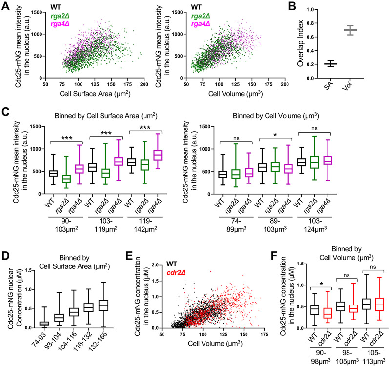
Figure 4. Cdc25 accumulation in the nucleus scales with cell volume. See also Figure S3 and S6.
(A) Cdc25-mNG nuclear mean intensity in WT, rga2Δ, and rga4Δ single cells plotted either by cell surface area (left) or by cell volume (right) (WT n=502, rga2Δ n=867, and rga4Δ n=684). The same cells are plotted in the two graphs. Experiment was repeated in triplicate and representative graphs from one experiment are shown. See also Figure S3A-C. (B) Overlap index analysis for data in panel A. SA, surface area. Vol, volume. (C) Cdc25 mean intensity in the nucleus for WT, rga2Δ, and rga4Δ cells binned by cell surface area (left) or volume (right). ***p<0.0001; *p<0.03; ns, not significant. n>90 for each strain per bin. Reported p value is the most significant value from pairwise comparisons within each group. (D) The concentration (μM) of nuclear Cdc25-mNG increases with cell size. Cells are binned by cell surface area (n>250 for each size bin). (E) Cdc25-mNG nuclear concentration (μM) in WT and cdr2Δ cells plotted by their cell volume (WT n=1,801; cdr2Δ n=1,037). (F) WT and cdr2Δ cells of the same volume have similar nuclear concentration (μM) of Cdc25 (n>110 per strain). Graphs show median as a line, quartiles, max, and min. *p=0.004; ns, not significant. See also Figures S1, S3, and S6.

