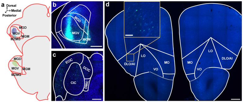
Figure 4. Retrograde tracer into MGN sparsely labels cell bodies in OFC.
a. Schematic of all injection sites in MGN (N = 3). Plate references from anterior to posterior: 34, 35 (Radtke-Schuller et al., 2016). Striped pattern denotes an injection that resulted in no observed cell body labeling in the OFC. b. Injection site of representative subject. Scale bar = 500 μm. c. In the same subject as B, expected labeling observed in IC. Scale bar = 500 μm. d. Representative OFC slice in the same subject as B. Scale bar = 500 μm, inset =100 μm.

