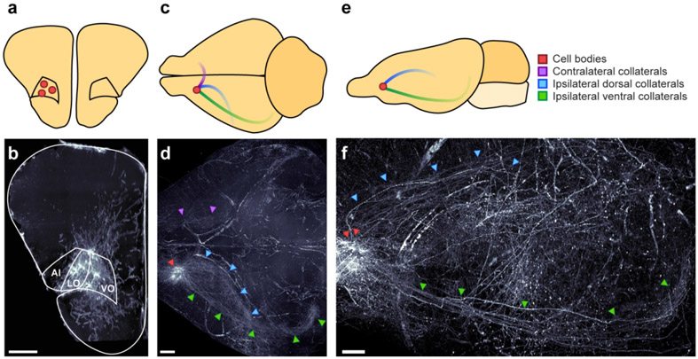
Figure 8. OFC neurons that project to auditory cortex send axon collaterals to other brain regions.
a-b. Coronal schematic (a) and section (b) depicting location of identified cell bodies in a representative subject. Section is a 175 μm thick coronal z-stack. Scale bar = 500 μm. c-d. Horizontal schematic (c) and view (d) depicting notable axon tracts emanating from OFC neurons in a representative subject. Scale bar = 1000 μm. e-f. Sagittal view (e) and section (f) depicting axon tracts in representative subject. Scale bar = 1000 μm.

