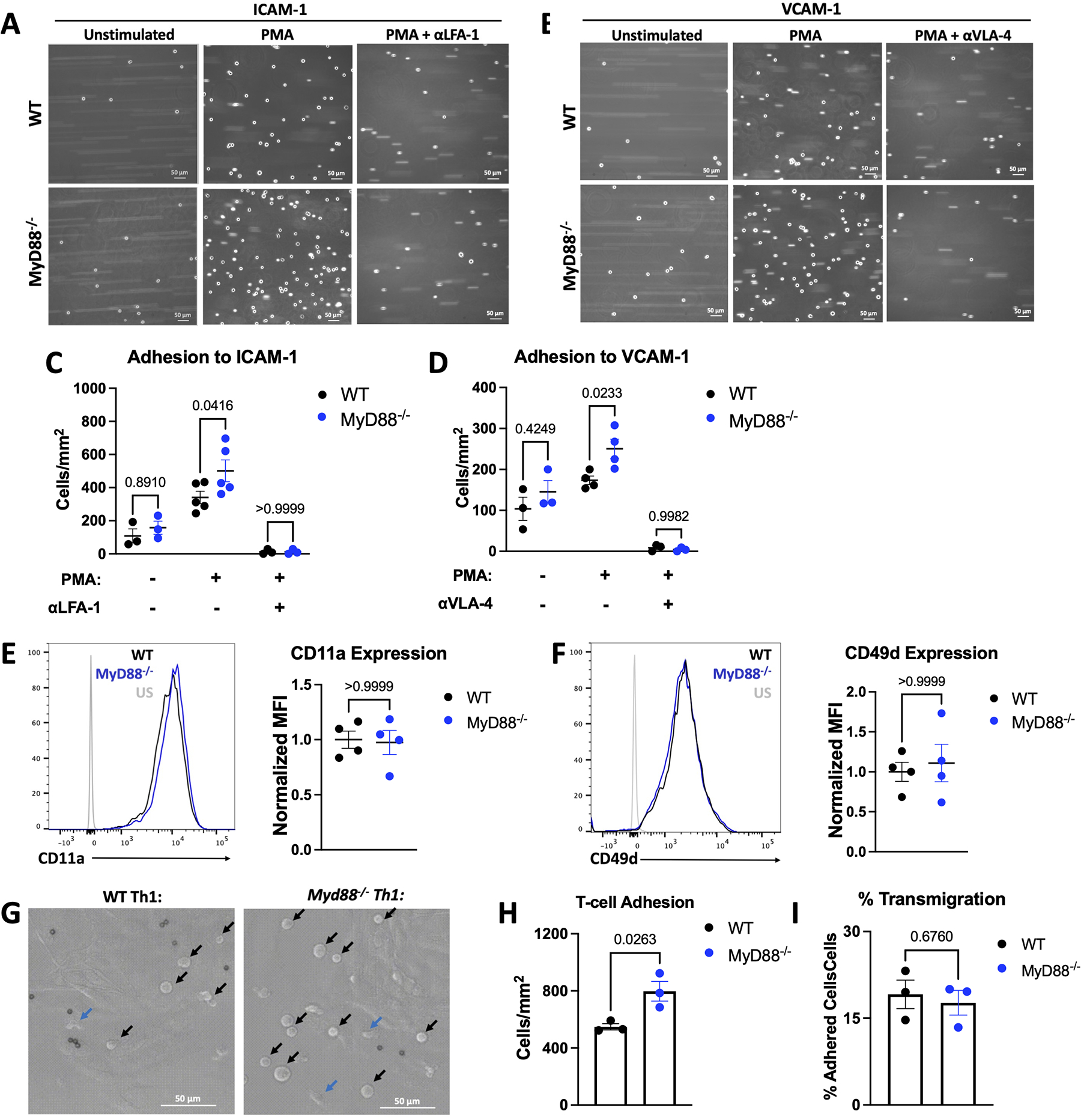
Figure 3: MyD88 Deletion Increases Th1 Adhesion Ability Without Impairment of Transendothelial Migration.

A-D. WT or Myd88−/− Th1 cells were perfused at 1 million cells / mL at an estimated shear stress of 1 dyne / cm2 over ICAM-1 or VCAM-1 after 5 minutes of treatment with 50 ng/mL PMA and/or 20 minutes of treatment with 20 μg/mL αLFA-1 or αVLA-4 where indicated, shown are representative images (A-B), quantified in C-D. E-F. Representative flow cytometry plots from WT or Myd88−/− Th1 cells shown with quantification of n=4 independent experiments. G. 1 million WT or Myd88−/− Th1 cells were perfused over mouse heart endothelial cells pre-treated with 125 ng/mL TNFα for 4 hours, at an estimated shear stress of 1 dyne / cm2, at 37°C. TEM was monitored over 10 minutes and adhered vs. migrated cells were counted manually. Representative images of adhered cells (black arrows) vs. transmigrated cells (blue arrows) in G. with quantification of adhesion in H. and percent migration (migrated / adhered) in I. In all panels each data point is from an independent experiment. All scale bars are 50 μM. Statistical analysis by 2-way ANOVA with Sidák’s multiple comparison test (C,D), Mann-Whitney Test (E, F) or T-test (H, I), p values shown.
