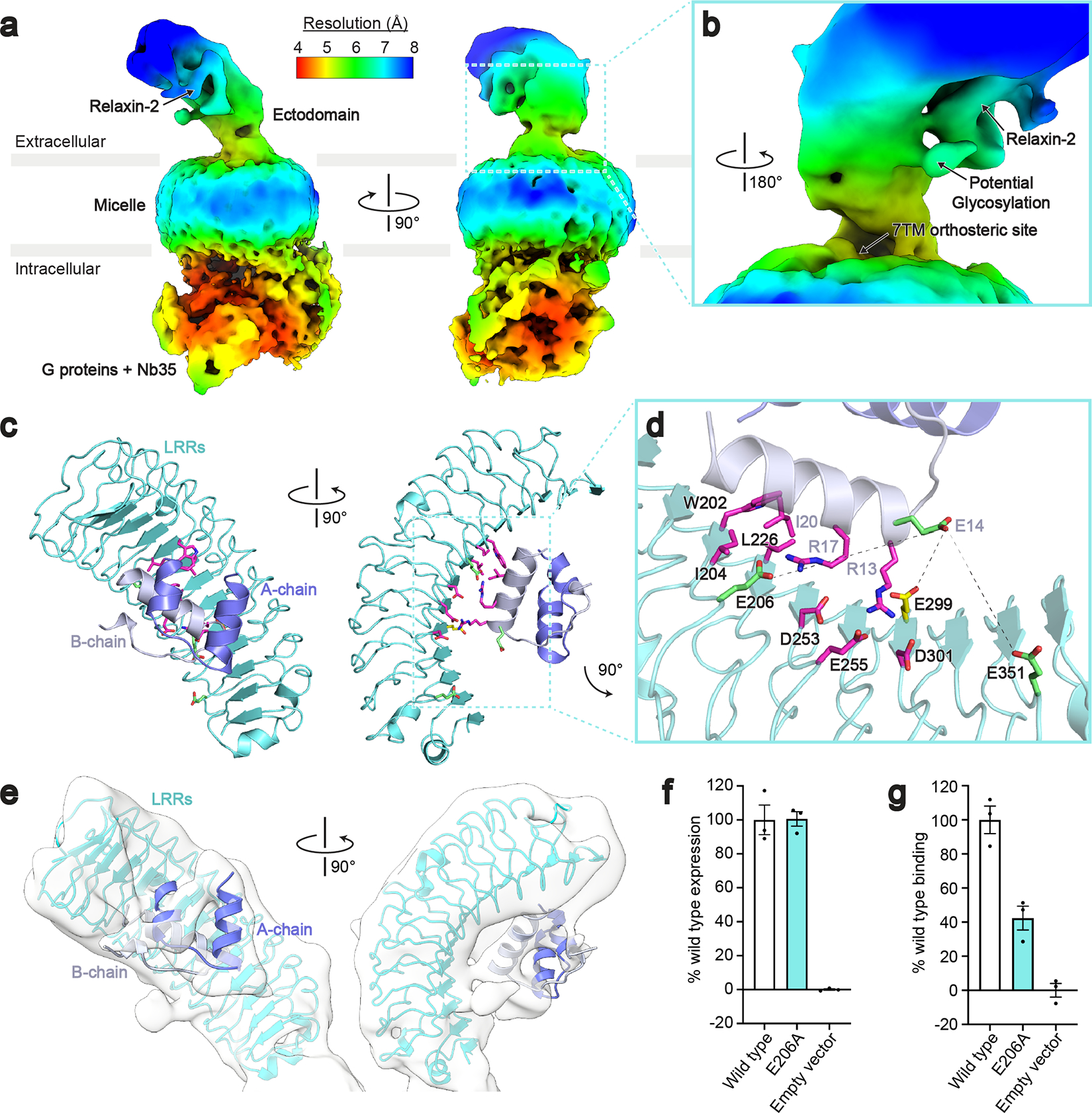
Fig. 4 |. Cryo-EM and crosslinking mass spectrometry reveal interactions between relaxin-2 and the leucine-rich repeats.

a, Local resolution cryo-EM map of the full-length RXFP1–Gs complex. b, The relaxin-2 binding site is above and rotated away from the 7TM orthosteric site. c, Model of the relaxin-2–LRR interaction from HADDOCK. d, Details of the relaxin-2–LRR interface with residues identified in published binding studies in magenta, residues from CLMS in green, and Glu299 from both CLMS and published binding studies in yellow. Crosslink distances: Glu14B-chain–Glu206 = 14.6 Å, Glu14B-chain–Glu299 = 10 Å, Glu14B-chain–Glu351 = 11.4 Å e, The relaxin-2–LRR model fit into the low resolution cryo-EM map. f-g, Receptor expression (f) and Fc-tagged relaxin-2 binding data (g) for the Glu206 to Ala mutation. Data are mean ± s.e.m., n = 3 technical replicates.
