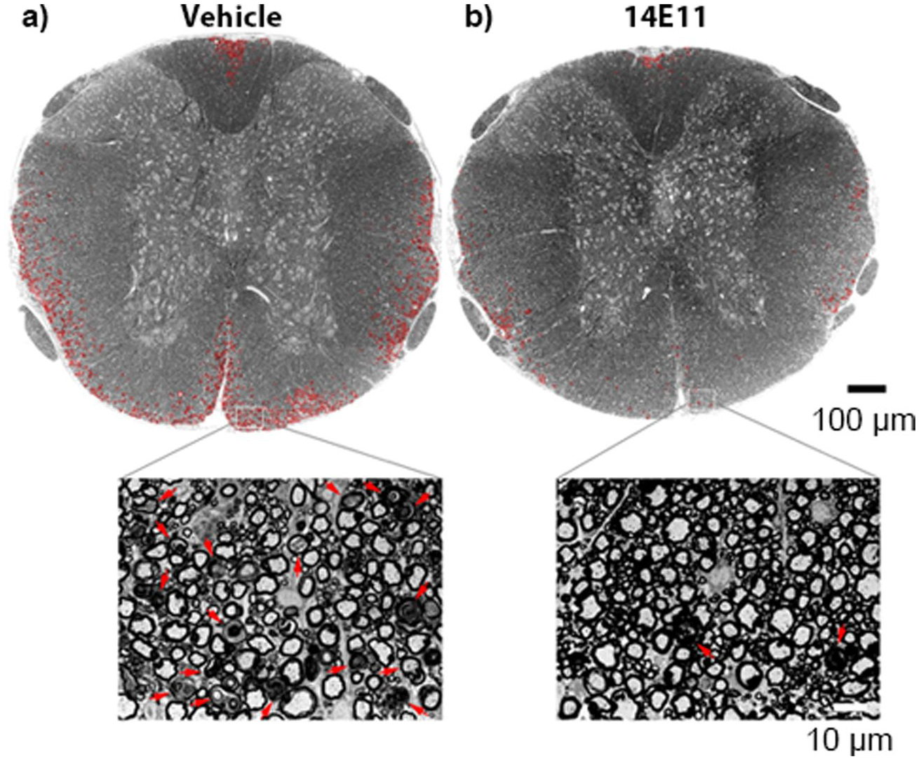
Fig. 3.

Axonal damage and demyelination in the thoracic spinal cord is reduced following treatment with 14E11. Damage to the myelin in spinal cord sections was assessed by staining the tissues with toluidine blue and using light microscopy to image the semithin sections at 20×. A complete view of the spinal cord cross section was obtained by manually stitching individual images into composites using Adobe Photoshop. The representative images shown are from mice treated with either vehicle (a) or 14E11 (b). Tissue damage in the white matter is denoted in red. The scale bar in the composite image indicates 100 μm, and the scale bar in the magnified view (63×) indicates 10 μm
