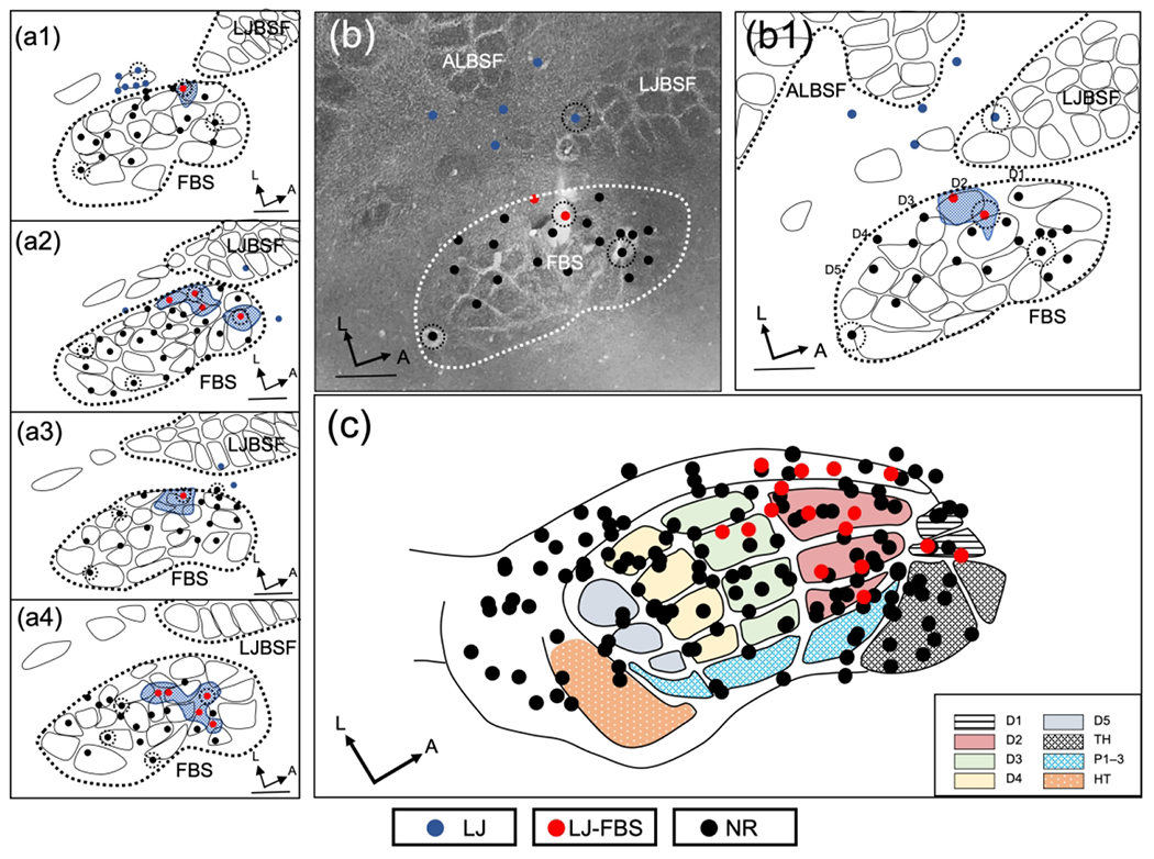
FIGURE 4.

Representation of lower jaw in the deafferented FBS – rapid brachial plexus anesthesia group (rBPA). (a1–a4) Representative examples of reconstructed physiological maps of four rBPA rats, mapped immediately after injection of lidocaine into the area of the brachial plexus. Lower jaw responsive sites in the deafferented FBS (LJ-FBS) are shown with red circles and the areas within the FBS enclosing these penetrations are shown with blue stippling. Lesion sites are designated with black-dashed circular lines around a recording site. Non-responsive recording sites (NR) where lower jaw responses were not found are designated by black circles; lower jaw responsive sites outside the FBS are designated with blue circles. (b) Photomicrograph of CO-stained section and (b1) accompanying line drawing reconstruction showing the location of new lower jaw responsive site in the deafferented FBS. (c) Responsive sites for all data collected in the FBS for seven rats are fitted to the FBS template; lower jaw responsive sites (red circles) and non-responsive sites (black circles). FBS nomenclature shown in box at lower right. Digit one (D1) through D5, thenar pad (TH), digit pads (P1–P3), hypothenar pad (HT), lower jaw (LJ), lower jaw recording sites in FBS (LJ-FBS), no-response (NR). Lower jaw barrel subfield: LJBSF; Anterior Lateral Barrel Subfield: ALBSF. Scale bars = 1 mm.
