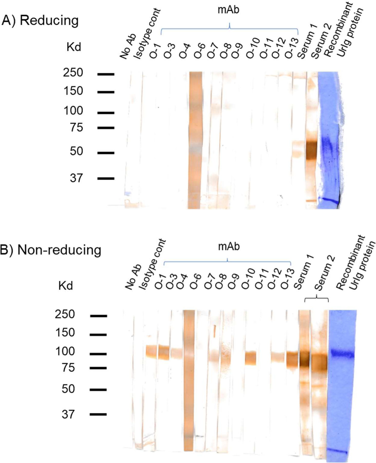
Fig. 7. UrIg monoclonal antibody specificity.

Western blotting test of UrIg-specific mAbs and antisera tested on recombinant UrIg protein under reducing (A) and nonreducing (B) conditions. The last lane on each blot is the UrIg protein stained by Coomassie Blue. All of the mAbs react with UrIg by ELISA (native), but only a subset (O-1, O-2, O-3, O-8, O-9, O-11, O-13) react with denatured and native UrIg. Antisera (from 2 mice) reacted with monomeric and dimeric UrIg, while the mAbs reacted only with the dimer.
