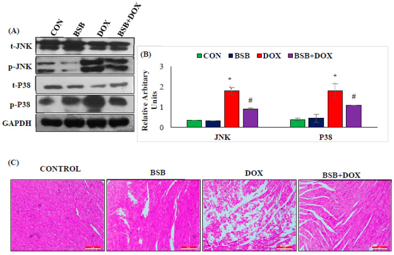
Figure 6.
Immunoblotting analysis of t-JNK, p-JNK, P38, and p-P38 (A), densitometric analysis and relative changes in the expression of JNK and P38 (B). Columns not sharing a common symbol (*, #) differ significantly from each other (* p < 0.05 vs. normal control, # p < 0.05 vs. DOX control). The histopathology of the myocardium (10×) (C). Normal control rat’s heart revealed a regular architecture of the myocardium; BSB-alone-treated rat’s heart also showed normal intact muscle fibers without any pathological changes; DOX-administered rat’s heart showed extensive muscle fiber degradation with inflammatory cells; BSB-treated DOX-injected rats showed reduced muscle fiber degradation without inflammatory cells. Histological analysis was performed in duplicate. CON: Normal control rats; BSB: α-Bisabolol-alone-treated rats; DOX: DOX-alone-treated rats; BSB + DOX: α-Bisabolol-and-DOX-treated rats.

